
Evidence date · 2026-03-06 · Research note 2 of 3
1 2 3
See current Track status →
Since the previous research note · Partly supported
Track continuity
Since the previous research note: When aging changes the immune neighborhood around a tumor
Previous expectation: Interrupting harmful senescence-related interactions around a tumor might improve checkpoint treatment, provided benefit and safety could be demonstrated in patients.
What this study adds: The current review gives this installment a specific immune-cell focus, including mouse-model accounts of BFAR inhibition restoring aged CD8 T-cell function and enhancing anti-PD-1 effects. Its account of tumor-killing senescence-like T cells also sharpens the distinction between removing cells and restoring their functions.
What changed in this study
An immune cell can become less able to multiply without losing every way it can attack a tumor. That apparent contradiction is central to a review published on March 6, 2026, about immunosenescence in cancer immunotherapy. It brings together findings on aging immune cells, tumor-driven dysfunction, and experimental ways to remove, protect, or restore them.
The treatment question is not simply whether an immune cell is “old” or “senescent.” It is which function has weakened, which functions remain, and whether the surrounding tumor has helped create the problem. A cell that expands poorly may be a poor source of reinforcements, yet still release substances that damage cancer cells. Removing it could relieve suppression in one setting and discard useful activity in another.
When an immune cell ages, one ability can fade before another
Chronological aging changes the T-cell pool. Naive T cells become less common, while terminally differentiated memory cells marked by loss of CD27 and CD28 become more prominent. These cells may show shortened telomeres, reduced telomerase activity, weaker signaling through molecules such as Lck and LAT, and higher levels of markers including CD57 and KLRG1.
Those features point to reduced renewal capacity, but they do not provide a complete description of what the cell can do. Multiplication and immediate cytotoxic activity are related but separable jobs. A T cell may be poor at producing a large new population while retaining, or even acquiring, an ability to kill targets through a different set of receptors and effector molecules.
Terminal differentiation is not identical to senescence, either. Nor is every change associated with aging proof that a cell has entered the same biological program. The review treats these categories as overlapping in some observations but not interchangeable in meaning.
Senescence and exhaustion are not the same state
T-cell exhaustion is usually discussed as a response to persistent stimulation, first characterized especially clearly in chronic viral infection. Exhausted cells often display inhibitory receptors such as PD-1, CTLA-4, Tim-3, and LAG-3, alongside impaired effector function.
Senescence is a broader stress response. The review describes it as arising from pressures such as replicative aging, DNA damage, or oncogenic stress, with features including cell-cycle arrest, resistance to apoptosis, and a senescence-associated secretory phenotype. A tumor can contain T cells showing aspects of both exhaustion and senescence, which helps explain why the terms are sometimes blurred in cancer research.
The difference matters for treatment. A therapy that blocks inhibitory signaling may not correct a cell whose limiting problem is durable loss of proliferative capacity. Conversely, removing a cell because it carries senescence-associated markers may overlook an immune function that has not disappeared. The marker is a clue to state, not a complete answer about therapeutic value.
Tumors can push tumor-directed T cells into a poor environment
The tumor microenvironment can help drive these changes. Tumor cells take up glucose and glutamine, while regulatory T cells can compete with effector T cells for glucose and maintain highly active glycolysis. Nutrient depletion and the accumulation of metabolites alter the conditions in which tumor-directed immune cells must work.
The review describes regulatory T cells as contributing to the induction of senescence in dendritic cells and T cells. Tumor cells can also promote senescence in transferred tumor-specific T cells. In melanoma models of adoptive immunotherapy, both the cancer cells and regulatory T cells were described as damaging the condition of the incoming cells.
This makes the environment part of the biological sequence rather than a backdrop. A T cell that enters a tumor may not behave like the same cell in a more supportive setting. Its reduced expansion can reflect the pressure of the tumor neighborhood, the cell’s prior history, or both. That distinction becomes important when considering whether to replace the cell, change the environment, or restore a function inside the cell.
Aged T cells may be poor expanders but active killers
One of the review’s more counterintuitive examples comes from aged mice. After laboratory activation, aged CD8 T cells showed senescence-like features but also displayed antigen-independent cytotoxic activity resembling that of natural killer cells. They had increased cytotoxic granules and higher expression of some natural-killer receptors.
The observation concerns what activated cells did in an experimental setting. A separate observation came from B16 melanoma models, where mice at least 30 weeks old showed less lung metastasis than young mice around 6 to 10 weeks old. That whole-animal result is consistent with the possibility that some aged effector CD8 T cells can contribute to antitumor defense through innate-like activity.
It does not mean that every senescent T cell remains useful, or that aging protects people from melanoma. It shows why proliferation and killing should not be treated as one measurement. A cell can be less capable of generating descendants while still releasing cytotoxic granules or responding through receptors that do not depend on the same antigen-recognition route.
Three treatment ideas target three different problems
The review separates three broad operations. Elimination removes senescent cells. Prevention or interruption blocks signals that push immune cells toward senescence. Restoration tries to recover useful activity from cells that have already become dysfunctional or aged.
Navitoclax illustrates the first approach in the review’s discussion. Eliminating senescent myeloid cells restored CD8 T-cell proliferation in vitro and overcame immunotherapy resistance in an in vivo setting. The review also cites a phase II study in chronic lymphocytic leukemia in which navitoclax combined with rituximab had better efficacy than rituximab alone. That clinical result belongs to its own disease and drug-combination context; it does not by itself show that removing senescent immune cells caused the benefit.
TLR8 agonism represents an attempt to prevent or interrupt dysfunction. Activating TLR8 on regulatory T-cell populations can disrupt the pathway by which they induce effector T-cell senescence, while TLR8 signaling may also reduce the ability of tumor cells to induce that state. The goal is not to clear a population after the damage is done, but to interfere with the local signals that create the problem.
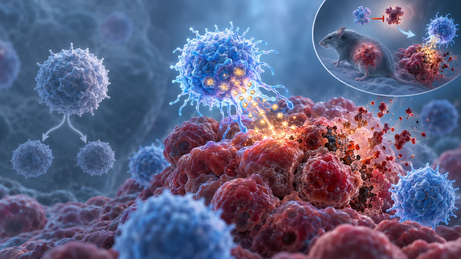
The BFAR example goes further toward restoration. In mouse tumor models, CD8 T cells from aged donors had difficulty becoming functional tumor-infiltrating resident memory cells. Removing BFAR genetically, or using the small-molecule inhibitor iBFAR2, restored antitumor activity in the aged T cells. iBFAR2 also enhanced the effects of anti-PD-1 treatment in those models.
That result describes a specific experimental route to recovering function in aged cells. It does not establish universal reversal of irreversible senescence, and it does not make senescence equivalent to exhaustion. Its importance is more precise: it suggests that at least some age-associated defects may be biologically actionable, and that restoring T-cell performance could complement checkpoint treatment in a tumor model.
Mouse and human aging do not line up neatly
Natural killer cells provide a warning against transferring an aging result from one species or tissue to another without adjustment. In older humans, the CD56bright NK-cell population becomes less responsive to interleukin-2 and less able to proliferate, while the more mature CD56dim population expands. Overall circulating cytotoxic capacity can appear relatively stable because there are more mature cells, even as cytotoxic function per cell declines through reduced activating-receptor expression.
Aged mice show a different pattern in most peripheral tissues. Their total NK-cell content and mature CD11b-positive, CD27-negative subset decrease, while early developmental stages in the bone marrow can remain similar. The pattern points toward impaired terminal maturation and reduced movement of mature cells out of the marrow.
The review discusses several possible reasons for the contrast, including differences in lifespan and NK-cell biology, laboratory environments that do not reproduce the immune history of people, and the heavy reliance on human blood samples rather than tissue measurements. The broader lesson is practical: a promising intervention aimed at an aging immune state may behave differently across species, cell compartments, and mechanisms. A drug combination that improves a clinical outcome also cannot be treated as proof of one proposed cellular pathway unless that pathway is directly demonstrated.
What this could change
A future checkpoint-treatment adjunct could be designed to restore tumor-fighting T-cell activity rather than simply remove cells carrying signs of aging. If the relevant mechanism holds up in patients, and if the intervention improves cancer control with acceptable toxicity, clinicians might eventually have a more selective way to preserve useful cytotoxic functions while reducing the immune suppression created inside tumors.
Reaching that possibility requires more than a favorable mouse experiment or a clinical combination result in a different setting. Patient studies would need to connect the targeted immune-cell state to the treatment mechanism, meaningful outcomes, and safety. The review’s contribution is to make that choice more concrete: the next generation of immunotherapy may need to decide whether a dysfunctional-looking immune cell should be cleared, protected from further damage, or given a chance to work again.
Key Sources
- Primary source: Dual roles of immunosenescence in cancer immunotherapy.
DOI or Journal Page · Review or Meta-Analysis · 2026-03-06