
Research Signal – Source date 2021-04-01
Older age was associated with worse COVID-19 outcomes, but the biology behind that pattern raised a difficult question. How can an immune system become less effective at recognizing and clearing a virus while also producing an excessive inflammatory response?
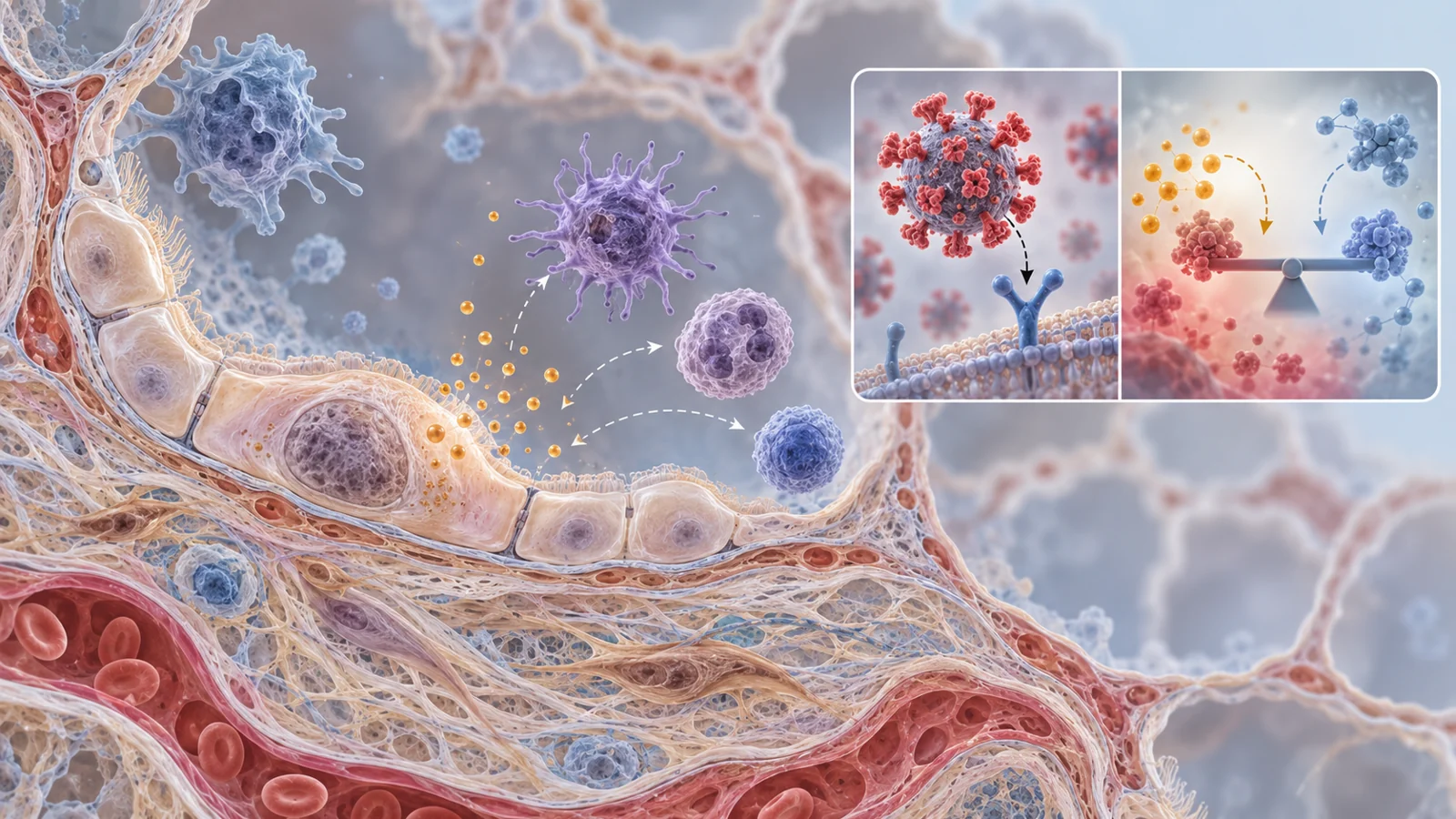
A review published on April 1, 2021, called An Account of Immune Senescence in the Clinical Pathophysiology of COVID-19 Infection in Aging, examines one possible answer. It places cellular senescence alongside broader age-related changes in immune function. Cells that have stopped dividing may continue releasing inflammatory signals, while aging immune defenses become less coordinated. The combination could help explain why protection weakens and tissue-damaging inflammation can still become intense.
The review presents this as a biological hypothesis, not a demonstrated explanation for age-related COVID-19 mortality. Its evidence combines observations from people with experiments involving other viruses and animal models, so the proposed chain must be followed carefully.
The puzzle of weaker defense and stronger inflammation
Aging changes the immune system in more than one direction. The review discusses a decline in lymphoid production from aging hematopoietic stem cells, with consequences for antigen recognition, memory CD8 T cells and responses to viral vaccination. At the same time, myeloid cells become more abundant but may be less effective at clearing a virus. A larger inflammatory response therefore does not necessarily mean a more capable antiviral response.
The other side of the puzzle involves the tissues themselves. Cellular senescence is a state in which a cell permanently stops proliferating. DNA damage, cellular stress and metabolic stress can push cells into this state. In younger or healthier tissue, immune surveillance can help remove some senescent cells. The review argues that this clearance becomes less effective with age, allowing senescent cells to accumulate.
Those two processes can coexist. Fewer effective lymphoid defenses may leave an older person less able to recognize and eliminate infection. Meanwhile, accumulated senescent cells may provide a persistent source of inflammatory signaling. The proposed connection is not that inflammation signals are inherently harmful; many are part of normal host defense. The concern is that a poorly controlled response may damage the same respiratory tissues that need to recover.
When a cell stops dividing but keeps signaling
Senescence describes a change in cell growth, not cellular inactivity. According to the review, senescent cells develop a senescence-associated secretory phenotype, or SASP. They release chemokines and cytokines including IL-6, IL-1β, TNF-α and other inflammatory mediators. Persistent DNA damage is discussed as one trigger for this secretory behavior.
This gives the hypothesis a possible mechanism. An older lung may contain more cells that no longer divide but continue sending inflammatory signals. A new viral infection could then add another layer of innate immune activation. The review connects this setting with reports of inflammatory cytokines and chemokines in people with COVID-19, including signals associated with severe disease and acute respiratory distress. It also notes that a major inflammatory surge may become detectable only after tissue injury is already developing, making the timing of intervention difficult.
The review further discusses the possibility that senescent lung cells provide a favorable environment for viral particle production because of their altered protein-synthesis activity. That proposal is part of the paper’s interpretation of the biology, not proof that senescent-cell burden determines the outcome of an individual infection.
The central uncertainty is causal. Older adults may have more senescent cells, weaker immune surveillance and greater vulnerability to severe disease, but those facts alone do not show that one factor drives the others. Senescence could be one contributor among several age-related changes, and the relevant burden may differ across tissues and patients.
ACE2 makes the story less simple
ACE2 adds a second layer of tension. SARS-CoV-2 uses the cell-surface protein as an entry point. The same protein also participates in the renin–angiotensin system by converting angiotensin II into angiotensin 1–7, a pathway the review describes as having anti-inflammatory effects.
The paper discusses findings suggesting that ACE2 expression declines with age. Viewed only through viral entry, less ACE2 might appear beneficial because there could be fewer gateways into cells. Viewed through inflammation-regulating signaling, however, reduced ACE2 could remove part of a system that counterbalances pro-inflammatory activity. The result is not a simple age-to-ACE2 rule in which more or less expression is automatically protective.
This matters for treatment ideas. A strategy aimed at blocking viral entry would need to account for ACE2’s other biological function. Similarly, a change observed in older tissue might reflect several interacting pathways rather than a single receptor effect. The review uses ACE2 to show why the biology of aging cannot be reduced to counting viral entry points.
The evidence changes with the virus and the model
The review’s reasoning becomes clearer when its evidence is separated by setting. Some observations concern COVID-19 patients, including reports of inflammatory mediators and severe respiratory complications. These observations describe what happens during human disease, but they do not identify which age-related process caused the deterioration.
Other examples come from different infections. In senescence-accelerated mice infected with influenza A virus, the review discusses reduced cellular immunity, greater viral persistence in the lungs and higher mortality. Those findings support the possibility that an aged or senescent biological environment can alter the response to respiratory infection. They do not directly test the same mechanism in older people with SARS-CoV-2.
The paper also discusses experiments involving SARS-CoV. Age-associated changes in Toll-like receptor signaling and fewer virus-specific CD8 T memory cells in aged mouse lungs are presented as possible contributors to worse lung pathology. MERS-CoV examples are used to discuss receptor biology and inflammatory signaling in senescent cells. Each experiment may illuminate a piece of the proposed mechanism, but the virus, host, tissue and experimental conditions are not interchangeable.
That is the main evidence-transfer problem. Results from influenza, SARS-CoV or MERS-CoV can motivate a hypothesis about SARS-CoV-2, yet they cannot by themselves establish that removing senescent cells would improve survival or reduce lung injury in older COVID-19 patients. The review brings these strands together to identify a research direction; it does not turn them into a clinical effect size.
Senescent-cell removal is not the same as changing autophagy
The therapeutic proposals in the review target different points in the suggested biology. Senolytics are compounds intended to remove senescent cells. If accumulated senescent cells are a meaningful source of damaging inflammatory signals, removing them could, in principle, change the tissue environment before or during infection. The supplied evidence does not show that this improves outcomes in older patients with COVID-19.
Autophagy is a separate process. It breaks down and recycles damaged cellular components during stress, infection and aging. The review notes that autophagy declines in aging and that viruses can manipulate the process. It discusses findings from coronavirus and other viral systems involving autophagy regulators, as well as compounds such as resveratrol, tomatidine, rapamycin, statins, carbamazepine and metformin as areas for investigation because of reported antiviral or autophagy-related activity in other contexts.
Changing autophagy is therefore not equivalent to removing senescent cells. One approach changes the population of cells in a tissue; the other changes an intracellular recycling and stress-response pathway. Either could affect more than the intended target. The review specifically highlights off-target effects and the need for in vitro and in vivo validation before candidate compounds could be judged for SARS-CoV-2 treatment.
A useful test would have to show more than an antiviral signal in a related system. It would need to establish whether the intervention acts on the proposed aging mechanism, preserves useful immune defenses and offers a favorable balance between benefit and harm in the patients whose biology it is meant to address.
What this could change
Aging biology could eventually become part of how severe COVID-19 is treated in older patients, rather than serving only as a broad risk category. Treatments designed to reduce damaging inflammatory activity from senescent tissue while retaining effective antiviral defenses could offer a more precise way to address the competing weaknesses of an aging immune system.
That possibility remains conditional. The implicated mechanisms would first need to prove causally relevant in SARS-CoV-2 infection, and interventions would need to demonstrate meaningful benefit without unacceptable off-target effects in the intended patients. The review’s value is to make that clinical possibility biologically imaginable while showing how much evidence must still connect the proposed mechanism to a safe treatment.
Key Sources
- Primary source: An Account of Immune Senescence in the Clinical Pathophysiology of COVID-19 Infection in Aging.
DOI or Journal Page · Review or Meta-Analysis · 2021-04-01
Keep exploring