
Research Signal – Source date 2021-01-12
The relationship between age and COVID-19 severity is not explained by a single failing immune switch. A review published on January 12, 2021, describes several age-related changes that can affect how the body recognizes and responds to coronavirus infections. Two ideas organize the discussion: immunosenescence, the gradual weakening and reshaping of immune function, and inflamm-aging, the tendency toward persistent age-associated inflammatory activity.
Together, they help explain an apparent contradiction. An older immune system may be less prepared to recognize a new virus quickly, yet more prone to produce inflammation that damages tissue once the response is underway. The review presents this as a biologically plausible way to understand vulnerability, not as one demonstrated causal chain that accounts for every severe case.
Two aging effects can coexist
A younger immune system generally contains a broader pool of naive immune cells. These cells have not yet encountered their matching antigen, so they provide more possible starting points for recognizing an unfamiliar pathogen. With age, the supply of new naive cells changes, while experienced and memory cells make up a larger share of the circulating population.
The balance matters because total cell numbers can look relatively preserved even as the range of possible new responses narrows. The review describes major age-related changes in the thymus, where T cells are produced. After thymic involution, production of naive T cells falls substantially. Peripheral cells can continue dividing to maintain overall T-cell numbers, but this replacement does not restore the same breadth of recognition for a novel antigen.
At the same time, aging is associated with a more inflammatory background. Immune cells and tissues may be more likely to maintain inflammatory activity, while the systems that coordinate and contain that activity become less well regulated. Existing illnesses common in later life can add to this burden. The result is not simply an immune system that is weaker in every respect. It is one whose timing, cellular composition, and inflammatory control have all changed.
Timing can shape the first antiviral response
The early response to a virus depends partly on type I interferons, antiviral signaling proteins that communicate through nearby and infected cells. When this signal arrives early, it can restrict viral replication and limit spread. When it is delayed, the virus may gain time while inflammatory processes continue to build.
The review discusses evidence that delayed type I interferon production increases with age and was observed in older individuals with SARS-CoV. That observation is relevant to the proposed biology, but it is not the same as a direct demonstration of the same timing pattern in older people infected with SARS-CoV-2. The distinction matters because the review brings together findings from related coronaviruses, human observations, and other experimental settings.
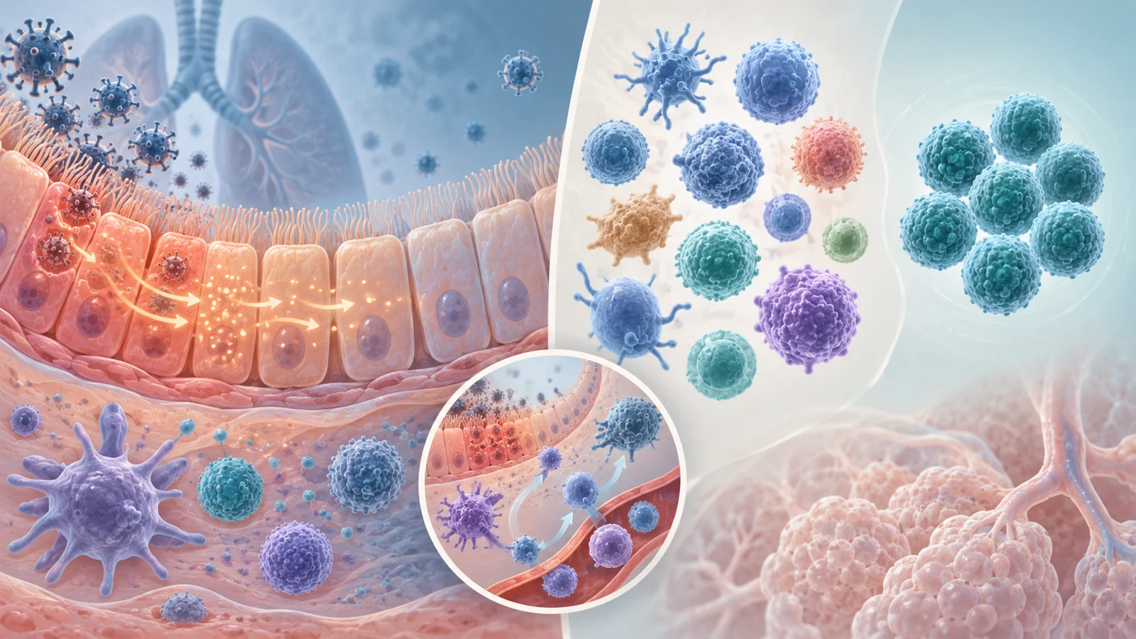
The later inflammatory sequence can also become harmful. SARS-CoV-2 infection of host cells can lead to cell injury and the release of danger signals. Nearby lung and vascular cells, along with macrophages and recruited immune cells, respond by producing inflammatory signals that attract more cells to the site. Recruitment can help control infection, but an inadequately regulated response may accumulate immune activity in the lungs and contribute to tissue injury, acute respiratory distress, and damage beyond the lungs.
Age may therefore affect both sides of the sequence. An early antiviral signal that is too slow can permit more viral activity, while an inflammatory response that is too persistent or poorly controlled can increase collateral injury. The review uses this timing problem to connect immune aging with disease progression, while leaving the contribution of each mechanism to individual outcomes unresolved.
Cell counts are not the whole adaptive story
Adaptive immunity depends not only on how many lymphocytes are present, but also on what they can recognize and how effectively they respond. The review notes that older adults may have lymphocyte levels similar to those of younger adults even though a greater proportion of their T cells have acquired experienced or memory-like characteristics. Those cells remain useful, especially against familiar or related antigens, but they do not provide the same starting repertoire as a large population of naive cells facing a new pathogen.
This helps clarify why a simple cell count can miss an important age-related change. A person may retain many T cells while having fewer uncommitted cells capable of rapidly generating a high-affinity response to an unfamiliar viral antigen. The review also discusses age-related changes in CD8 T-cell function, including findings from animal work in which older cells showed less proliferation or responsiveness after stimulation.
Memory cells are also central to vaccination. After an immune encounter, some antigen-specific T and B cells persist and can respond more rapidly if a similar antigen appears again. Tissue-resident memory cells remain in sites such as the lungs and may provide local protection, but the review describes age-related changes in these cells as well. In older settings, increased or altered tissue-resident memory populations were associated in experimental work with more inflammatory responses.
The practical implication is not that vaccination has one fixed result in every older adult. It is that age can influence the cellular starting point, the quality of recognition, and the persistence of immune memory. Those are biological reasons to study vaccine strategies with attention to older immune systems rather than assuming that the response of a younger adult is the default.
What the comparisons can and cannot tell us
The review does not rely on one type of study. It discusses human observations from COVID-19, earlier coronavirus research, aged-mouse experiments, studies of immune cells, and reports involving potential treatments. These sources can illuminate different parts of the problem, but they do not all answer the same question.
For example, aged mice showed higher expression of some exhaustion-associated markers on CD8 cells and reduced ability to proliferate after antigen stimulation. In a study of SARS-CoV infection in monkeys, older animals had lower CD8 counts and later developed higher viral levels than younger animals. Such findings offer mechanistic clues about how aging might affect antiviral control. They do not by themselves establish that the same cellular changes, with the same importance, occur in older humans with SARS-CoV-2.
The review also discusses regulatory T cells, which help restrain inflammatory immune cells. One reported observation linked lower levels of naive regulatory T cells with greater COVID-19 severity. The authors explicitly note that the relationship’s causal role was unknown. A correlation can arise because disease changes immune-cell levels, because another factor affects both, or because the cells contribute to disease. The observation identifies a relationship worth investigating, not a confirmed lever for changing outcomes.
Age also travels with other conditions. Diabetes, hypertension, chronic lung disease, cardiovascular disease, and obesity were associated in the review with more severe COVID-19, and several are common among older adults. Age-related immune changes and accompanying illnesses may interact, but the selected material does not assign a precise independent contribution to each one. This is why the review is most useful as a guide to connected biological questions rather than as a calculator of an individual person’s risk.
Why an inflammatory target is not yet a treatment answer
Inflammation can point researchers toward a treatment idea without proving that the target is the cause of the worst outcomes. The review highlights interleukin-6, or IL-6, a cytokine involved in immune-cell trafficking and inflammation. Higher serum IL-6 was associated with more severe COVID-19 and mortality in the observations discussed, while elevated IL-6 is also part of the inflammatory profile associated with aging.
That combination can make IL-6 look like a plausible point for intervention. Yet a marker can be a driver, a consequence, or part of a larger process. Blocking it could help in one context, do little in another, or interfere with useful immune activity depending on timing and the patient’s overall condition.
The review gives siltuximab as a concrete example. In a compassionate-use report involving 21 people with COVID-19-related acute respiratory distress syndrome, the review reports that after seven days, 33% had improved, 43% were stable, and 24% had worsened. One death and one cerebrovascular event were also reported. Because the experience had no control group, the patient trajectories cannot show whether siltuximab caused the improvements, whether some patients would have improved without it, or how the treatment affected the reported adverse events.
The lesson is narrower and more useful than a treatment recommendation. Age-related inflammatory biology can generate testable therapeutic hypotheses, but an attractive mechanism and a small uncontrolled clinical experience are not the same as demonstrated benefit.
What this could change
Better understanding of immune aging could eventually support therapies and vaccination strategies adapted to the way older adults recognize viruses, produce antiviral signals, and regulate inflammation. The downstream goal would be better protection against severe infection, with choices shaped around the older immune system rather than a younger person’s response assumed to be universal. That benefit remains conditional on the proposed mechanisms translating into interventions that show meaningful benefit and acceptable harms in older people.
Key Sources
- Primary source: Aging, Immunity, and COVID-19: How Age Influences the Host Immune Response to Coronavirus Infections?
DOI or Journal Page · Peer-Reviewed Paper · 2021-01-12
Keep exploring