
Research Signal – Source date 2026-04-08
Aging can weaken the immune system’s ability to respond to new threats while leaving inflammatory activity simmering. That combination may seem contradictory. It is central to a review published on 8 April 2026 that examines how age-related immune changes could interact with brain aging and neurodegenerative diseases.
The review brings together work on Alzheimer’s disease, Parkinson’s disease, amyotrophic lateral sclerosis (ALS) and multiple sclerosis (MS). Its proposed connection runs through several interacting parts: immune cells that become less responsive, senescent cells that release inflammatory signals, changes in the blood–brain barrier, and brain support cells whose protective work can falter. The evidence includes experimental models and human observations, which answer different questions about cause and treatment.
A weaker response can coexist with persistent inflammation
Immunosenescence means age-related changes that reduce or alter immune function. Inflammaging is the persistent, usually low-grade inflammation associated with aging. Neither term means that every older person has the same immune state, or that ordinary aging is itself a neurodegenerative disease.
Aging immune cells may become less effective at recognizing threats, presenting antigens and mounting adaptive responses. Yet damage signals and other persistent stress cues can continue to activate innate immune cells. Those cells may release inflammatory messengers such as IL-6, TNF-α and IL-1β. The result can be poor defense alongside ongoing inflammation: the response is less well coordinated, but it does not simply switch off.
Senescent cells can help sustain this pattern. These cells have stopped dividing, but may remain active and release a mixture of inflammatory substances called the senescence-associated secretory phenotype, or SASP. When immune clearance becomes less effective, senescent cells can accumulate. Their secretions can, in turn, promote pathways such as NLRP3 inflammasome activation and cGAS–STING signaling, adding to inflammatory activity. This creates a feedback loop: weaker clearance permits more senescent cells to persist, and their signals add to the burden the aging immune system must manage.
The review distinguishes this background of immune aging from disease-associated amplification. In neurodegenerative conditions, protein aggregates and other disease-specific processes can intensify inflammation or impair its resolution. Immune aging may make the brain more vulnerable to those pressures, while the disease process can further alter immune function. The proposed relationship is therefore two-way, rather than a claim that age-related immune changes alone initiate these illnesses.
The brain’s support cells have protective work to do
Microglia are immune cells that reside in the brain. In healthy conditions, they help survey the tissue and clear debris and protein aggregates. Astrocytes support neurons and synapses, help maintain the brain’s local environment, and interact with microglia. These activities are part of ordinary brain maintenance, not evidence of disease by themselves.
With age, microglia can show features associated with cellular senescence, including altered metabolism, excess reactive oxygen species, impaired phagocytosis and release of SASP factors. Phagocytosis is the process of taking up and removing material. The review describes reduced CD36 signaling and inhibitory CD22-related signaling among contributors to weaker clearance of cellular debris, myelin debris and aggregates such as amyloid-β and α-synuclein. When cleanup becomes less efficient, material that should be removed can remain in the tissue, while inflammatory signals persist.
Astrocytes also undergo changes that can affect brain support. The review describes losses in proteostasis, or the cell’s ability to maintain and manage proteins, alongside lysosomal and proteasomal dysfunction, impaired support for synapse formation, and accumulation of autophagosomes. Astrocyte and microglial function are interdependent: changes in astrocytes, including altered IL-10 receptor signaling and cholesterol biosynthesis, may also affect microglial homeostasis.
Mitochondria connect cellular stress with immune signaling. The review describes defective autophagy and mitophagy, the processes that help clear unwanted cellular material and damaged mitochondria, as contributors to microglial senescence. Mitochondrial dysfunction can increase oxidative stress; release of mitochondrial DNA can activate cGAS–STING signaling and help sustain inflammation. These mechanisms are described across experimental and disease contexts. They suggest ways that cellular stress and inflammatory activity might reinforce one another, while leaving the extent and timing of those interactions in people unresolved.
A changing barrier alters traffic and cleanup
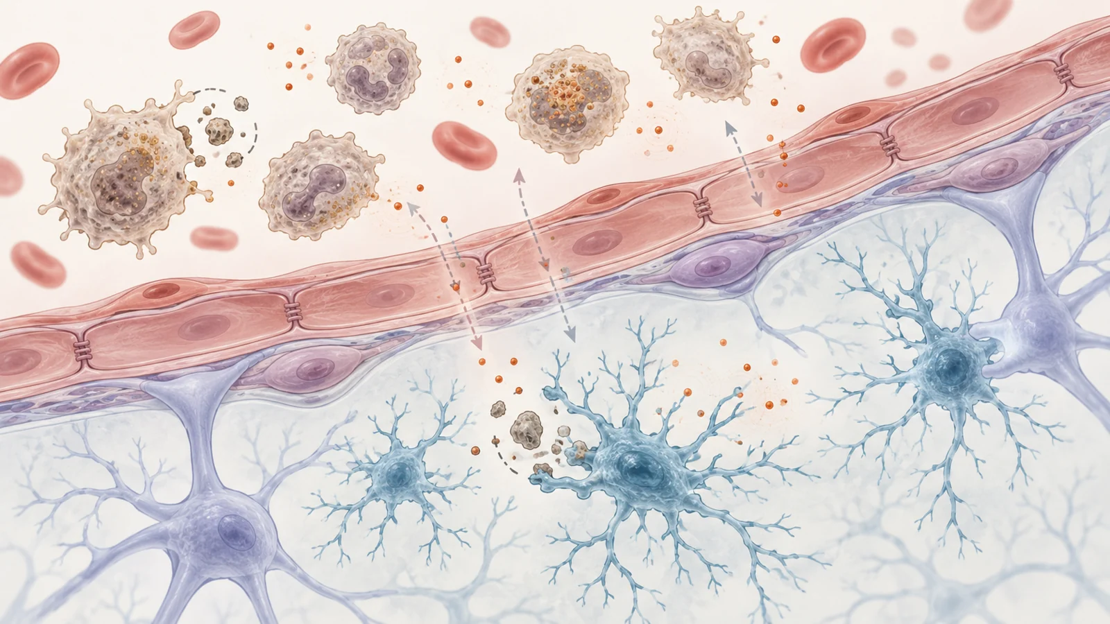
The blood–brain barrier is a regulated interface between the blood and brain tissue. It helps control what passes into the brain and supports the movement of some material out. It is not a sealed wall: its cells and surrounding structures respond to signals from both sides.
The review describes age-related changes to this interface, including disrupted tight junctions, altered transport across cells, thickening of the basement membrane, endothelial mitochondrial dysfunction and loss of pericytes. Pericytes help support blood-vessel function. Together, these changes may allow more circulating cytokines, chemokines and immune cells to enter brain tissue, linking the state of the peripheral immune system with local inflammation. The review also notes that barrier deterioration can occur during cognitively healthy aging, so the change alone does not identify a neurodegenerative disease.
The barrier may affect protein clearance as well as immune traffic. The review describes lower activity of the efflux transporters P-glycoprotein and LRP1, which help move amyloid-β out of the brain, and higher activity of RAGE, a receptor that can carry circulating amyloid-β in the other direction. Microglial activation, changes in astrocyte endfeet and pericyte loss may also weaken barrier integrity. This route is distinct from the systemic senescent-cell feedback loop: it concerns the interface through which signals, cells and proteins move between blood and brain. In combination, altered traffic and less effective clearance could add to conditions that favor neuroinflammation.
Immune cells can protect as well as inflame
Adaptive immune cells do not have a single role in the aging brain. Senescent-like CD8+ T cells, for example, have been associated with cognitive decline. In experimental models described by the review, these cells can enter the brain in part through CXCL10/CXCR3 signaling and produce interferon gamma (IFNγ). The resulting inflammatory environment can affect oligodendrocytes, the cells that support myelin, as well as neurons and neural stem cells. In some models, removing CD8+ T cells reduced age-associated oligodendrocyte loss; stronger CD8+ activity or added IFNγ worsened measures of glial activation and neurodegeneration.
Regulatory T cells, or Tregs, normally help restrain immune activity and maintain balance. Aging can impair their frequency or function, weakening that restraint. The effect of T-cell aging therefore depends on which cells are involved and what they are doing: some may contribute to inflammatory injury, while others help limit it.
B cells also resist a simple “more or less immunity” interpretation. In an Alzheimer’s disease model, non-amyloid-specific antibodies enhanced plaque dynamics by supporting microglial phagocytosis through Fc receptors. In another experimental finding discussed by the review, removing adaptive lymphocytes worsened pathology, while restoring them mitigated neuroinflammation. These model results point to protective and harmful functions within adaptive immunity. Broadly suppressing immune activity could remove useful functions along with damaging ones; the challenge is to understand which responses, in which compartments, matter in a given disease.
Four diseases, different immune contexts
The review uses several diseases to illustrate that immune aging meets distinct biology in each condition. In Alzheimer’s disease, microglia normally take up soluble and aggregated amyloid-β through receptor-mediated processes. Age-related changes that weaken clearance may intersect with amyloid accumulation, oxidative stress and glial inflammation. The proposed connection here centers on protein handling and brain-resident immune cells.
In Parkinson’s disease, the relevant disease features include loss of dopamine-producing neurons and aggregation of α-synuclein. The review describes changes in peripheral immune-cell populations, including fewer naïve cells and more late-differentiated or senescent T-cell profiles. Some profiles are associated with disease measures such as dopamine-transporter levels or age at onset. These observations connect immune aging with Parkinson’s disease, but do not show that one immune profile starts the disease.
For ALS, the review highlights associations between faster progression and increased senescent or late-memory T and B cells in peripheral immune profiling. Experimental evidence also links CD4+ T-cell senescence with neuroinflammation and motor-neuron degeneration. This places adaptive immune changes alongside the disease’s progressive motor-neuron loss, while human causality remains unsettled.
MS presents a different context: chronic autoimmune neuroinflammation, with signs of premature immune aging. The review describes thymic involution, telomere shortening, expansion of late-differentiated CD8+ T cells, accumulation of age-associated B cells and accelerated epigenetic aging in peripheral immune cells. These changes are associated with greater disability, reduced response to disease-modifying therapies and a higher risk of transition to progressive MS. Here, immune aging intersects with an already immune-mediated disease process rather than serving as a single explanation for neurodegeneration.
What the evidence can and cannot tell us
Some causal evidence comes from experimental models. Manipulating immune cells or using senolytic compounds to remove senescent cells has altered inflammation and disease-related outcomes in animals. Such experiments can test whether a pathway contributes to a process under controlled conditions. They do not establish that the same intervention will be effective, safe or appropriately targeted in people.
Human evidence described in the review is mainly observational. Higher levels of inflammatory markers such as IL-6, TNF-α and C-reactive protein have been associated with faster cognitive decline and dementia risk. Longitudinal studies summarized by the review link higher midlife IL-6 and CRP with later cognitive deterioration and hippocampal atrophy. These patterns support a biologically plausible connection, but an association does not show that a marker caused the change. It also does not make the marker a validated clinical test or show that lowering it would improve an individual’s outcome.
The findings span different diseases, immune compartments and experimental settings. That variety is useful for identifying possible shared mechanisms, but it limits how confidently any one result can be carried across conditions. The review describes immune aging as a contributor and amplifier worth investigating, while human causal evidence and proven disease-modifying treatments remain limited.
What immune aging could change for brain treatment
If researchers can identify which aging immune changes actively worsen a particular disease, and target them without disrupting protective immune functions, future treatments might preserve brain-cell support or improve the clearance of harmful material. Senolytics, immune-modulating approaches, dietary strategies and microbiome-based interventions are among the directions the review discusses. Most remain preclinical or at an early clinical stage, and the review reports limited evidence of disease-modifying benefit in people. The potential practical gain is a more precise way to protect brain function in specific disease contexts; reaching it depends on establishing who might benefit, when to intervene and how to do so safely.
Key Sources
- Primary source: Immunosenescence and Inflammaging as Drivers of Neurodegeneration: Cellular Mechanisms, Neuroimmune Crosstalk, and Therapeutic Implications.
DOI or Journal Page · Review or Meta-Analysis · 2026-04-08
Keep exploring