
Research Signal – Source date 2025-11-27
Aging changes the immune system as well as the brain. Could those immune changes help drive Alzheimer’s and Parkinson’s disease, or do they mostly accompany damage already underway? A review published on November 27, 2025, brings together evidence on both possibilities and proposes 11 hallmarks to organize research on immune aging, also called immunosenescence.
The review’s contribution is a way to connect changes across the body with changes at the brain’s borders and inside brain tissue. It also surveys possible treatments aimed at those changes. Its evidence comes from different kinds of prior work, including human associations, cell studies, animal models and intervention research. The paper is not a new clinical trial showing that reversing immune aging slows Alzheimer’s or Parkinson’s disease.
Aging reshapes the immune system across several sites
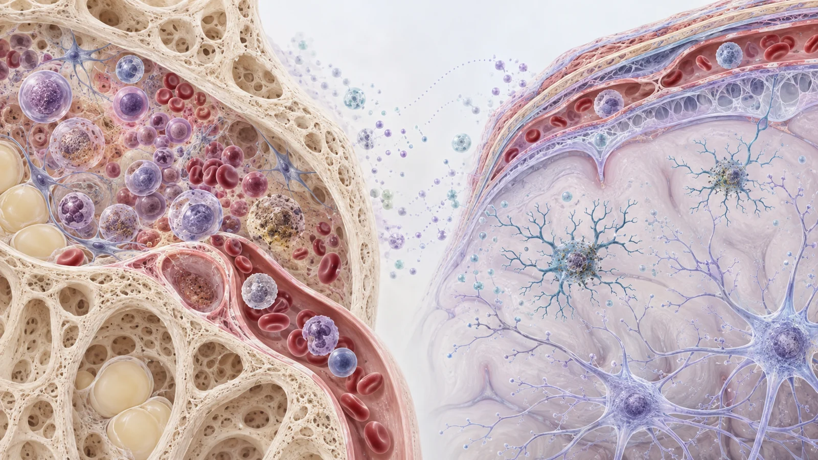
Some changes begin in bone marrow, where blood and immune cells develop. With age, hematopoietic stem cells—the cells that replenish blood-cell populations—lose some self-renewal and regenerative capacity. The balance of their output also shifts: myeloid cells such as neutrophils and monocytes become more prominent relative to lymphoid cells. Meanwhile, the marrow has fewer naïve T cells and fewer B-cell precursors, alongside an accumulation of senescent CD8 T cells, a population marked in part by loss of the CD28 surface protein.
These shifts matter because immune cells do not act in one place. Cells in blood and other tissues can influence the brain, while immune populations at the brain’s borders and within its tissue have their own local roles. The meninges, protective layers surrounding the brain, contain T cells and antibody-producing cells. In older mice, some antigen-experienced B cells can move from the blood into the meninges, and the number of IgA-producing plasma cells near dural venous sinuses increases with age. At the same time, aging is associated with reduced drainage through meningeal lymphatic vessels, a change that may contribute to the buildup of misfolded proteins in the central nervous system.
Inside the brain, microglia are resident immune cells that help monitor and clear material. With age, some microglia accumulate lipid droplets and show impaired phagocytosis—the process of engulfing and clearing material—along with excess reactive oxygen species and inflammatory signals. Myelin fragments released as the brain ages can be taken up by microglia; the review describes how this burden may contribute to lysosomal dysfunction and further weaken cleanup. These observations offer a possible connection between immune aging and the brain’s capacity to maintain its tissue environment.
The authors group such changes under 11 proposed hallmarks: genomic instability, telomere attrition, epigenetic dysregulation, stem cell exhaustion, loss of proteostasis, deregulated nutrient-sensing, mitochondrial dysfunction, cellular senescence, chronic inflammation, altered intercellular communication and microbiome dysbiosis. The categories overlap. A cell with accumulated damage, for example, may function less well and release inflammatory signals that affect neighboring cells. The value of the list is to organize a broad set of possible mechanisms; it does not mean every hallmark has been demonstrated in every immune cell or brain disease.
Senescence can connect inflammation with impaired cleanup
Cellular senescence is a lasting state in which a cell stops dividing after stress or damage. Senescent cells can release a mixture of inflammatory molecules known as the senescence-associated secretory phenotype, or SASP. In the brain, the review describes senescent microglia and astrocytes as potential sources of signals that damage nearby neurons and intensify neuroinflammation. Immune-cell senescence may also contribute to a wider cycle in which inflammation and tissue dysfunction reinforce one another.
Recognizing senescence is not straightforward. Researchers look for combinations of signs, including cell-cycle arrest, DNA-damage responses, SASP, increased lysosomal content and changes in cell structure or survival pathways. But immune cells are diverse, and some markers overlap with other cell states. Surface markers such as CD57, KLRG1 and NKG2D, or a loss of CD28, can help identify particular senescent immune populations; no single marker by itself resolves the question for every cell type.
This measurement challenge matters for interpreting the broader framework. A label such as “senescent” needs to be tied to the cell being studied and the evidence used to identify it. The review’s central proposal is that these cellular states and other aging-related processes could help explain how immune function changes with age, including in the brain. Whether the same mechanisms operate in peripheral blood cells and brain-resident immune cells remains an open research question.
Alzheimer’s and Parkinson’s offer different kinds of clues
For Alzheimer’s disease, the review discusses human observations linking immune-cell features with disease and cognition. People with Alzheimer’s have been reported to show shorter telomeres in peripheral blood mononuclear cells than age-matched controls. In one reported association, T-cell telomere length—but not B-cell or monocyte telomere length—tracked with global cognition scores. Other studies described increases in certain age-associated CD8 T-cell populations, including cells with low levels of the IL-7 receptor and cells called TEMRA, which have been considered senescent. These populations were associated with cognitive measures.
Such findings make blood immune profiles plausible candidates for studying disease progression, but they are associations. They do not by themselves show whether immune aging contributes to cognitive decline, reflects other disease-related changes, or participates in both directions.
The Parkinson’s evidence highlighted in the review includes experimental work on alpha-synuclein, a protein central to Parkinson’s pathology. In mouse models, alpha-synuclein pathology was linked to senescence-like changes in microglia, including inflammatory signaling, mitochondrial dysfunction and expression of senescence-associated markers. Alpha-synuclein-containing extracellular vesicles from disease models also increased PD-L1 expression in T cells in experimental settings, a change linked to suppression of those cells. These results suggest routes by which disease-related protein pathology might alter immune cells. They do not yet explain how those changes relate to symptoms or disease course in people.
The relationship may run both ways. Aged microglia and astrocytes may be less able to handle abnormal protein buildup, while proteins such as amyloid beta, tau and alpha-synuclein can affect immune cells. The review notes that whether these proteins directly accelerate aging in peripheral immune cells is still poorly understood. So the evidence points to possible feedback between disease pathology and immune aging, while leaving the direction and importance of that feedback unresolved.
Treatment results vary by disease and study
The review surveys 12 categories of proposed approaches, including rejuvenating stem cells, clearing senescent cells, reducing inflammation, supporting mitochondrial function and changing nutrient-sensing pathways. The evidence ranges from theoretical proposals to preclinical experiments and some clinical studies. These categories are a research agenda, not a single tested treatment package.
Several Alzheimer’s mouse-model studies described in the review reported improvements after interventions aimed at senescent cells or immune aging. In one tauopathy model, genetic clearance of senescent cells reduced gliosis, tau-related changes and neurodegeneration. In a 5xFAD mouse model, a senolytic drug improved cognition and reduced brain inflammation. Other experiments reported that transferring young bone marrow or immune cells into Alzheimer’s-model mice reduced plaque burden or inflammation and improved behavioral outcomes. Results in these models are useful for testing mechanisms, but they do not establish benefit in patients.
A different result came from a multiple-sclerosis model. Neither a senolytic drug combination nor genetic clearance of senescent cells improved disease severity in experimental autoimmune encephalomyelitis. That negative finding cautions against assuming that senescent-cell removal will help across neurological diseases; the relevant cells and disease processes may differ.
Human findings on spermidine, a compound discussed in relation to protein maintenance and autophagy, also point in different directions. One study reported improved memory performance in older adults at risk of dementia, while a randomized trial in people with subjective cognitive decline found no memory benefit compared with placebo. The contrast illustrates why an intervention’s biological rationale and one positive study do not settle whether it will help a particular group of patients.
The brain-specific causal question remains
A major gap is whether the hallmarks used to describe immune aging in peripheral cells also explain aging in microglia and other brain-associated immune cells. Do these cells experience the same kinds of telomere change, mitochondrial dysfunction and epigenetic shifts, or do their local environment and tasks produce different aging pathways? The review argues that answering this requires studying brain immune cells directly rather than assuming that findings from blood apply unchanged.
A related question is causal: which immune changes, if any, help initiate or sustain neurodegeneration, and which arise in response to it? Human associations and experimental models illuminate different parts of that problem. The review brings them into a shared account, but they do not establish one causal pathway that applies across Alzheimer’s, Parkinson’s and other neurodegenerative diseases. Nor does its treatment survey demonstrate a broadly effective immune-rejuvenation therapy for patients.
Could immune-cell treatments protect the aging brain?
A better account of brain-specific immune aging could eventually help clinicians match treatment to the immune-cell pathway contributing to an individual’s disease. If researchers can identify causal targets in the brain, alter them safely and show that doing so preserves function in patients, immune-directed treatments might become one way to protect brain health. That possibility depends on connecting cell-level changes to meaningful patient benefit; the review sets out the questions and candidate strategies, not a proven route to that outcome.
Key Sources
- Primary source: Immunosenescence in aging and neurodegenerative diseases: evidence, key hallmarks, and therapeutic implications.
DOI or Journal Page · Review or Meta-Analysis · 2025-11-27
Keep exploring