
Research Signal – Source date 2025-07-19
Fat is often treated as stored fuel, but it also helps regulate metabolism and immune activity. Its location and condition change with age, raising a consequential question: can those changes help drive problems in other organs, or do they mainly reflect aging elsewhere?
A July 2025 review brings together evidence that points to several plausible routes from fat tissue to the rest of the body. It describes changes in storage capacity, tissue structure, and chemical signaling, alongside early shifts observed in mouse studies and associations found in human tissue. The evidence makes fat a candidate contributor to systemic aging. It does not yet show that fat changes start the process in people.
Fat has different jobs in different places
White adipose tissue is the body’s main energy store. Its cells take up excess energy and hold it as triglycerides, then release that stored energy when needed. Brown fat, which is much less abundant in adults than in newborns, can dissipate energy as heat. Beige fat is also thermogenic; it is distinguished as a separate functional category in mice.
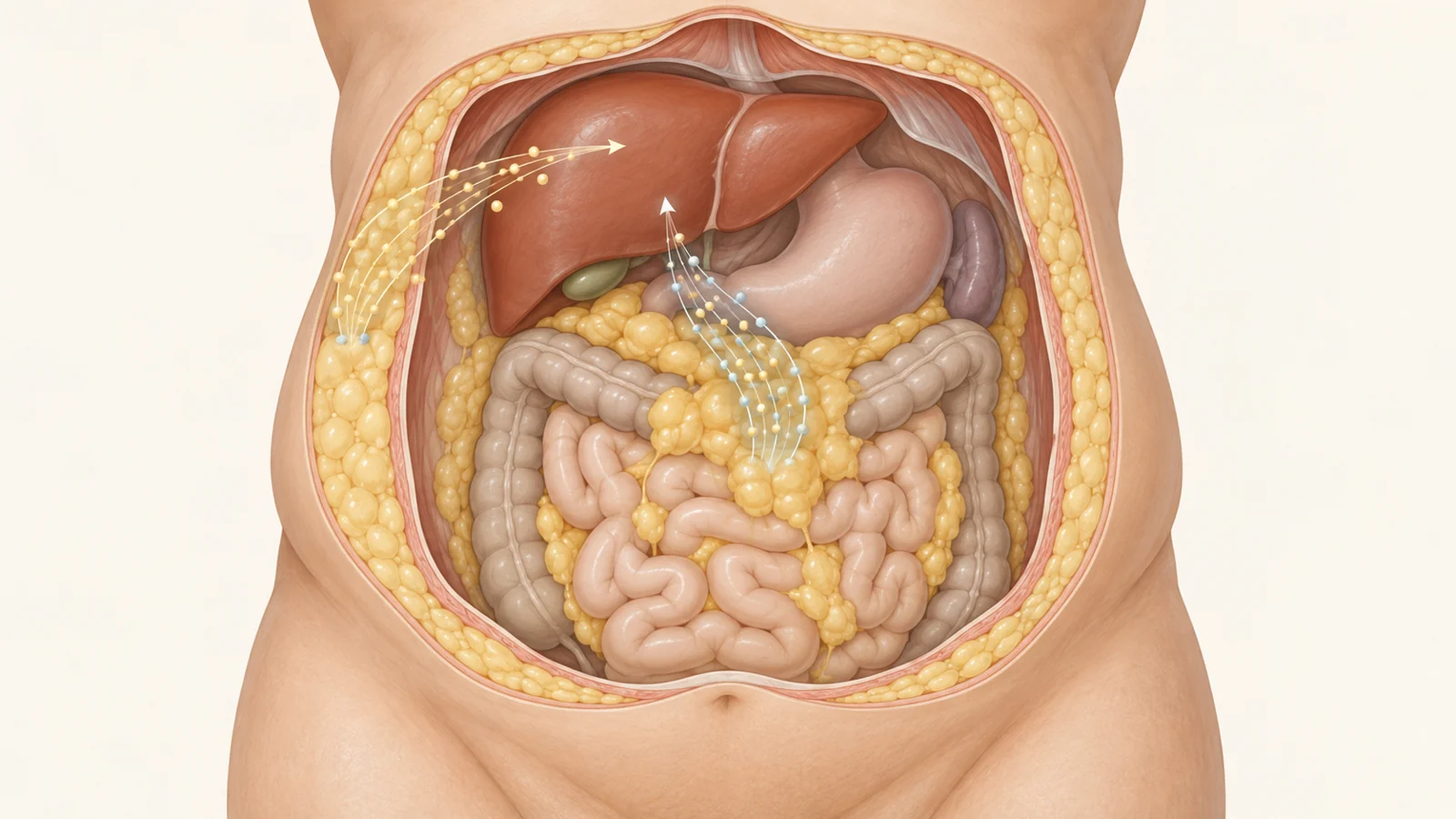
These tissues are not interchangeable. Fat depots differ in their origins and in their metabolic, endocrine, and heat-producing properties. With age, the pattern of fat can change: subcutaneous fat may shrink, while visceral fat around internal organs expands. Fat may also accumulate in places such as the liver, heart muscle, and skeletal muscle.
A body-fat total cannot show these shifts. Nor does it say whether the remaining tissue can safely store incoming lipids, release them when needed, or maintain its other functions. The location and condition of fat matter alongside its amount.
When storage space becomes less flexible
As fat cells enlarge, they can store more lipid, which may help keep excess fat from reaching other organs. But enlargement has costs. A larger cell has less surface area relative to its volume, which can hinder nutrient transport and cell signaling. The review also describes enlarged fat cells as releasing more inflammatory factors, including IL-6 and TNF-α.
The surrounding tissue can become less accommodating, too. Aging fat is associated with fibrosis, in which collagen and other material build up in the tissue’s supporting structure. In aged mice, the review cites a study that found a sevenfold increase in collagen staining in visceral fat. It also describes a possible link between restricted oxygen supply and fibrosis: oxygen-related signaling can promote the production and cross-linking of collagen. The sequence and causal contribution of low oxygen remain uncertain, but fibrosis is associated with a less flexible tissue environment.
Together, impaired storage and reduced flexibility offer one way fat dysfunction could affect distant organs. If fat tissue cannot buffer lipids effectively, more may accumulate outside fat cells, where excess lipid can harm tissues. The review connects this possibility with ectopic fat in the liver and the progression of metabolic dysfunction-associated fatty liver disease. This is a proposed route, not proof that aging fat initiates disease in people.
Fat also changes the messages it sends
Lipid spillover is only part of the story. Adipose tissue secretes adipokines and other molecules that help regulate energy use and immune activity. The review describes age-related shifts in these secretions, including lower adiponectin and higher leptin, and notes that a lower adiponectin-to-leptin ratio is associated with measures such as body mass index, waist circumference, and insulin levels. It also cautions that direct evidence connecting leptin to fat aging remains limited.
The immune environment within fat changes as well. Studies in mice report more T cells in visceral fat with age, and a shift in macrophages toward a more inflammatory profile. Senescent cells can add to local inflammation by releasing a mixture of signaling molecules. In one human tissue analysis discussed in the review, a pro-inflammatory group of adipocyte progenitor cells was more abundant in samples from older people.
These observations suggest a second route by which aging fat could influence distant tissues: altered secretions and immune signals may affect organs beyond the depot where they arise. But findings about particular cells, molecules, and tissues do not by themselves show how much each contributes to disease across the human body.
Early changes are not a human timeline
The review’s timing argument draws on evidence from different settings. In mouse aging analyses, white fat showed early changes: metabolism-related cell populations declined during an earlier age interval, while immune-cell populations expanded more markedly later. In another mouse experiment, a short high-fat-diet challenge triggered inflammation in fat without a similar response in other metabolic tissues; prolonged feeding was associated with stronger inflammatory responses in the liver and muscle.
These models address different questions. An age-profile study tracks changes across life in mice. A diet challenge tests what happens under a particular excess-energy exposure. Neither establishes that fat is the first cause of systemic aging in humans.
Human tissue observations add relevance, but they provide a different kind of evidence. Analyses of fat samples have found age-related inflammatory features, and some studies associate measures of fat aging with metabolic measures. Such links can identify patterns worth investigating; they cannot determine whether tissue changes came before altered glucose metabolism, followed it, or developed alongside it. The review calls for longitudinal studies to clarify how fat dysfunction unfolds over time.
Images and biopsies capture different clues
Imaging can show where fat is distributed. CT and MRI are described as reference methods for measuring the balance between subcutaneous and visceral fat, while DXA and ultrasound may offer alternatives for broader or repeated assessment. These methods address anatomy and distribution; they do not directly reveal the cellular state of a depot.
A biopsy samples tissue itself. The review discusses staining for senescence-associated beta-galactosidase activity and evaluating fibrosis as ways to examine fat aging more directly. In a prospective study of 227 people with obesity, measures from subcutaneous and omental fat were associated with glucose-metabolism abnormalities and differences in fat distribution. That association makes tissue sampling informative, but it does not reveal which change came first. Imaging and biopsy therefore answer complementary questions, rather than providing one complete measure of fat aging.
What intervention studies have tested
The proposed role of fat in systemic aging has prompted several kinds of intervention research, but their endpoints should not be treated as interchangeable.
In aged mice, low-dose treatment with a PPARγ-activating drug altered gene expression in visceral fat and was associated with less inflammation and fibrosis, as well as improved tissue homeostasis. Those results suggest that changing fat function may be possible in an animal model. They do not establish that the same approach improves health in older people.
Senolytics are designed to remove senescent cells. The review describes promising mouse findings involving insulin sensitivity, fat inflammation, and metabolic balance. The human trials it discusses involved other conditions, not treatments aimed directly at senescent cells in adipose tissue. They therefore offer limited evidence about feasibility in people, not proof of an adipose-specific benefit.
Dietary calorie restriction has also been studied in several ways. A two-year randomized trial in older adults reduced subcutaneous, visceral, and intermuscular fat. Separate research cited by the review links longer-term restriction with changes in inflammatory and senescence-related measures in fat. Changes in fat volume, however, are not the same endpoint as restoring fat function, and neither by itself demonstrates longer human healthspan. For senolytics in particular, the review calls for human trials that directly target adipose senescence and assess effects on metabolic health.
What healthier fat could mean for other organs
If fat tissue could retain the capacity to store lipids and regulate its secreted signals with age, it might help protect organs such as the liver from some metabolic strain. That possibility depends on a causal link between fat dysfunction and downstream harm, and on interventions that can improve fat tissue in people and demonstrate a tissue-specific benefit. Longitudinal human evidence is needed to show how these changes unfold and whether improving fat function can make a meaningful difference beyond the tissue itself.
Key Sources
- Primary source: Fat talks first: how adipose tissue sets the pace of aging?
DOI or Journal Page · Review or Meta-Analysis · 2025-07-19
Keep exploring