
Research Signal – Source date 2026-03-31
Cancer becomes more common with advancing age. That makes treating the biology of aging an appealing possibility for cancer prevention and care. But some changes associated with aging also stop damaged cells from multiplying. Removing those barriers could work against cancer protection.
A scientific synthesis published March 31, 2026, brings this tension into focus. In Cancer and aging: complex associations and therapeutic targets, the authors organize nine shared biological processes according to whether they encourage cancer, restrain it, or act differently depending on the setting. Drawing together existing research, they explore the prospect of treating cancer and aging-related dysfunction through shared targets. The drug examples span cells, mice, and patients, with different outcomes measured at each level.
How damaged DNA changes both cells and tissues
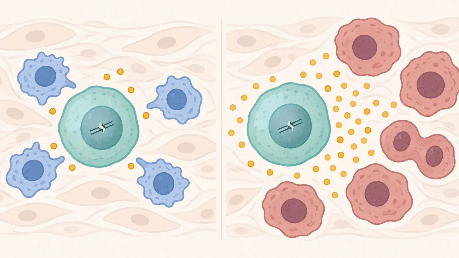
DNA damage creates two problems that can develop alongside each other. As repair becomes less effective with age, persistent damage can activate responses that stop cells from dividing. Stem cells, which help replenish tissues, can become depleted. The result is less capacity to replace lost or damaged cells and maintain tissue function.
Other cells acquire mutations that give them a growth advantage. Changes can activate genes that promote proliferation or disable genes that normally suppress tumors. Successive rounds of growth and selection can then favor increasingly malignant populations. The same broad failure to maintain DNA can therefore contribute to declining tissue renewal and to the expansion of particular damaged cells.
The surrounding tissue helps determine which cells thrive. The authors describe aging tissues in which the material around cells becomes stiffer, oxygen and nutrients become unevenly available, and immune activity shifts toward persistent inflammation. These conditions alter the pressures under which cell populations compete. A mutation’s consequences unfold within that changing environment.
This adds an important layer to the familiar idea that cancer reflects mutations accumulated over time. A cell carrying a growth-promoting alteration also encounters signals from immune cells, neighboring cells, and the surrounding tissue. The authors connect those interactions to changes in metabolism and communication between cells, explaining how damage at one level can reshape conditions at another.
When stopping cell division protects the tissue
Cellular senescence is a state of sustained cell-division arrest. A senescent cell remains biologically active even though it has stopped multiplying. That arrest can prevent a damaged or potentially malignant cell from producing a growing population of descendants.
Shortening telomeres provide one concrete route into this state. Telomeres are protective structures at chromosome ends. In most ordinary body cells, they shorten with repeated divisions because the DNA-copying machinery cannot fully reproduce those ends. When they become critically short, a damage response can impose a growth barrier.
Some cancer cells escape that barrier by acquiring ways to maintain their chromosome ends. One route involves reactivating telomerase, an enzyme that adds telomeric DNA. This helps explain why a process associated with diminished tissue renewal can also limit uncontrolled growth—and why restoring a cell’s ability to keep dividing is not automatically a desirable outcome.
Arrested cells also communicate with their neighbors. Their released molecules are collectively called the senescence-associated secretory phenotype, or SASP. During an acute response, these signals can recruit immune cells to help eliminate cells at risk of becoming malignant. If inflammatory secretion persists, however, it can supply growth and survival signals to nearby cancer cells. The authors discuss interleukin-6 and other signaling molecules as part of this process.
The distinction is between what happens inside the arrested cell and what its continued activity does to the tissue around it. Growth arrest can remain a barrier to that cell’s proliferation while persistent secretions support other cells. This is why counting senescent cells alone cannot explain all their consequences; their signals and neighboring cell populations also matter.
Cancer treatment can add another source of injury
The relationship also runs in the opposite direction. Cancer and its treatment can contribute to processes associated with functional decline.
The authors describe how chemotherapy can damage DNA in normal tissues, disrupt mitochondria, and increase reactive oxygen molecules. Radiation can produce persistent oxidative stress and genomic damage through overlapping mechanisms. These injuries can trigger therapy-induced senescence, leaving cells in sustained growth arrest after treatment.
The synthesis also discusses how prolonged, intense immune activation during immune-based treatment may contribute to exhaustion and broader changes in immune function. These are pathways through which injury can occur; their consequences vary across treatments and patients.
For someone receiving cancer care, this connection reaches beyond the immediate tumor response. Persistent tissue dysfunction can affect quality of life and contribute to late complications. The therapeutic question becomes whether cancer control can be accompanied by less lasting injury to the tissues that patients depend on during recovery and afterward.
What the drug examples actually measured
The authors describe a “one drug, two targets” ambition: act on a shared pathway to address both cancer and aging-related dysfunction. Three examples show how much the meaning of a promising result changes with the experimental setting.
In cellular models, the outcome is a change in cells. The synthesis reports that dasatinib combined with ellagic acid produced synergistic clearance of senescent cells. It also discusses dasatinib with resveratrol increasing programmed cell death in senescent human lung fibroblasts, cells involved in tissue structure and repair. These findings concern the response of particular cell populations to particular combinations.
Such experiments allow researchers to examine whether senescent cells can be removed and how their survival changes. The measured unit is the cell population. Translating that effect into a benefit for an intact tissue requires understanding what happens to the other cells and functions that depend on it.
In mice, the aging-related and cancer findings come from separate studies. A table entry describes an antibody called RMP1-14, directed at the immune-regulating protein PD-1. One cited mouse study reported improvements in aging-related kidney and liver features. Another reported improved survival in mice with diffuse large B-cell lymphoma.
The distinction matters because these outcomes were not jointly demonstrated in the same animals. The table brings together an organ-related result in one experimental setting and a cancer-survival result in another. Their shared intervention makes the pairing scientifically interesting, while each study answers its own biological question.
In patients, the example concerns a defined blood cancer treatment setting. The authors discuss adding navitoclax to ruxolitinib in people with myelofibrosis whose response to ruxolitinib alone was suboptimal. Myelofibrosis involves disease of the blood-forming tissue and scarring in the bone marrow. Navitoclax appears in the synthesis because of its senescent-cell clearance activity and its potential against blood cancers.
In the phase II findings summarized by the authors, 26.5% of patients achieved at least a 35% reduction in spleen volume at week 24. At that time point, 30% achieved at least a 50% reduction in total symptom score. Separately, 33% of evaluable patients showed a one- to two-grade improvement in bone marrow fibrosis. These percentages describe different endpoints and should not be added together as an overall response rate.
Anemia and thrombocytopenia—low red blood cell and platelet levels—were also observed. The authors describe these adverse events as manageable, but they remain part of interpreting the combination’s clinical effects. The findings concern spleen size, symptoms, and marrow scarring in a specific disease setting. They do not measure reversal of aging across the body.
Why these findings do not yet add up to a general treatment
The three examples address different questions. Clearing senescent cells in a cellular model, changing organ features in mice, and improving disease-specific outcomes in patients are distinct achievements. Putting them together does not establish that a single intervention simultaneously extends healthy human lifespan and improves cancer outcomes. That conclusion requires evidence connecting both benefits within an appropriate clinical setting.
There is also no simple scale on which less biological aging always means less cancer. The authors highlight telomere biology as one example: short telomeres can be associated with cancer risk, yet exceptionally long telomeres can also raise the risk of several cancers. The effects of a biological feature cannot be inferred from whether it looks younger or older in isolation.
Selective action is therefore a central challenge for treatment development. Removing a harmful cell population, changing its inflammatory output, and restoring a cell’s proliferation are different interventions. They need to be evaluated for their effects on normal tissue, malignant cells, and the wider immune environment. Success against one cellular feature cannot stand in for evidence of overall patient benefit.
What this could change
If interventions against shared pathways can reduce persistent treatment-related tissue dysfunction while maintaining tumor control, cancer care could better protect the capacities people need after treatment. For older patients in particular, that could mean retaining more physical function and independence, with fewer lasting complications and better quality of life.
The causal path is concrete: effective treatment controls the malignancy, while a selective intervention limits damaging cellular persistence or inflammatory signaling in normal tissues. To make that approach useful, clinical studies would need to demonstrate those patient benefits without weakening cancer control or introducing unacceptable toxicity.
The prospect is cancer care that takes longer-term recovery into account alongside the tumor response. The synthesis gives researchers biological reasons to pursue that goal and shows why the resulting treatments must be judged by how well patients live as well as how their cancers respond.
Key Sources
- Primary source: Cancer and aging: complex associations and therapeutic targets.
DOI or Journal Page · Peer-Reviewed Paper · 2026-03-31
Keep exploring