
Research Signal – Source date 2026-05-27
A tissue has a chronological age: the time since the animal or person was born. Its gene activity can tell a more layered story. In a study spanning four mammals, researchers built transcriptomic clocks—measures based on gene activity—that estimated chronological age and mortality-related change. The two kinds of readings did not always respond alike.
The distinction matters because a clock is a model, not a direct measurement of how long an individual has left to live. The researchers trained one set of models to estimate age and another using mortality-related targets. Those models could therefore register different features of the same biological state, including changes associated with health and lifespan in the studied animals.
What the clocks learned from gene activity
The team combined 11,165 tissue samples from mouse, rat, macaque and human, drawn from more than 25 tissues. The dataset brought together newly generated mouse data and published gene-expression measurements. The researchers used it to build models for chronological age and for two related measures: normalized age and expected mortality.
For the rodent mortality targets, the researchers used survival data to fit Gompertz models for experimental groups, including different interventions. These models estimated expected mortality at a given age and group; they did not observe an individual animal’s future lifespan. A normalized-age measure also placed chronological age in relation to the group’s expected maximum lifespan. The resulting targets let the clocks learn patterns linked to both ageing and lifespan differences in those models.
The expression data came from separate studies, where technical differences can make samples difficult to compare. To reduce that problem, the researchers expressed gene activity as a change relative to age-, sex- and strain-matched control samples from the same dataset and tissue. In everyday terms, the models learned whether a sample’s gene activity sat above or below a local reference, rather than treating raw measurements from different studies as directly interchangeable.
That choice mattered for testing the models beyond the samples used to build them. In tests that held out entire datasets, the relative multi-tissue clocks retained predictive ability. When tissues were held out, chronological-age clocks still captured age patterns across those tissues, with median correlations around 0.86 to 0.88. In tests leaving out one species at a time, the chronological clock tracked age in the unseen species with correlations from 0.66 to 0.84. These results point to shared patterns within the mammals and tissues tested, while leaving open how well the models would transfer beyond them.
Shared patterns appeared across cells and species
The analysis also asked whether these broad patterns were visible within individual cell types. In mouse cell-type data, 48 of 49 types showed a significant positive relationship between chronological age and the age-clock reading; 46 of 49 did so for the mortality clock. A separate comparison of sorted cells from young and old mice also found age-related differences in each cell type with enough samples for analysis. Stem-cell populations were among those showing shifts associated with mortality-oriented readings.
Those findings suggest that the changes captured by the clocks were not confined to one organ or to mature cells. They do not mean every cell ages in the same way. Rather, the researchers found recurring expression patterns across many tested tissues and cell types, alongside pathway-specific differences that become easier to see when the model is broken into components.
Pathway clocks separated different responses
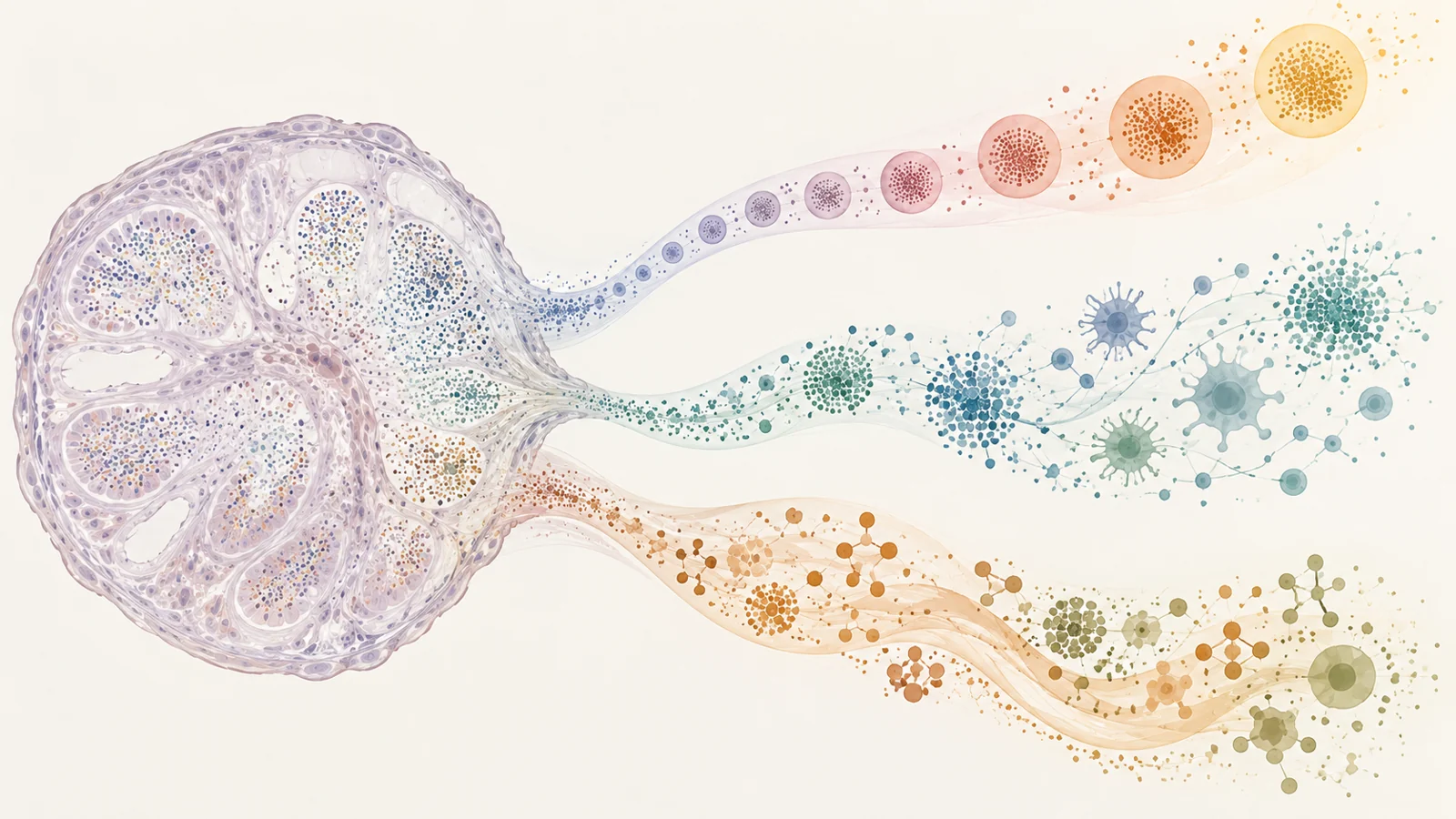
To interpret what drove the overall readings, the researchers grouped genes that tended to change together into modules. A module is a set of co-regulated genes associated with a cellular function or process. The team developed clocks for 23 modules, including inflammation, interferon signalling, mitochondrial function, chromatin modification and extracellular matrix organization. Together, the module genes captured much of the mortality-related variation in the larger model, though each individual module clock was less accurate than a clock using many genes at once.
The module readings could respond differently to the same broad category of challenge. In a mouse model of acute inflammatory stress, the strongest mortality-related shifts appeared in immune modules, including inflammation and interferon signalling. In liver samples from calorically restricted mice, readings shifted across several modules, with particularly notable changes in metabolism-related groups such as lipid metabolism and oxidative phosphorylation. These are distinct patterns: an overall molecular-age estimate can combine them, while module clocks can show which systems contributed most strongly in a given setting.
Cellular reprogramming and metabolic stress offered another contrast. In induced pluripotent stem cells, most chronological and mortality clocks showed lower readings, with the strongest shift in the extracellular-matrix and cell-identity-related module. By comparison, metabolic stressors increased mortality-oriented readings mainly in metabolic modules. These experimental findings show that gene-expression patterns can move in different directions across pathways under different conditions. They do not establish that an intervention reverses human ageing or extends human life.
A tumor exposed the limits of one combined number
A liver-tumor result illustrates why a composite clock needs interpretation alongside its parts. The tumors had a lower overall mortality-related reading than age-matched controls. On its own, that could sound like a younger profile. But the component patterns were mixed: cell-cycle activity, dedifferentiation and extracellular-matrix changes contributed to the lower reading, while inflammatory and metabolic signals moved in an ageing-associated direction.
A rapidly changing tissue can therefore combine features that a single summary number compresses. In this case, proliferation and shifts in cell identity coexisted with inflammation and metabolic disruption. The result is not evidence that the tumors were biologically healthier or that cancer reduces ageing. It shows how a composite estimate can be shaped by distinct processes, and why pathway-level readings are needed to understand what sits behind it.
Human findings linked gene activity to outcomes
The study tested its mortality clocks in human blood data from the Framingham Heart Study. After adjustment for age and sex, readings from a universal composite clock, a multi-module clock and several individual module clocks were associated with all-cause mortality. The module associations included chromatin modification, inflammation and oxidative phosphorylation or haem metabolism. Several transcriptomic clocks performed comparably to second-generation DNA-methylation clocks in these analyses, and the best chronological-age clock was not necessarily the strongest mortality predictor.
This is an association between a blood-based reading and subsequent mortality in the study population. It does not make the clock an individual countdown or show that using it to make clinical decisions improves outcomes. It also illustrates why accuracy at estimating birthdays and association with mortality are different performance questions: the gene-activity features that closely track age need not be the features most associated with later death.
A separate analysis looked at proteins in blood plasma from more than 50,000 UK Biobank participants. Higher levels of GPNMB, CDKN1A and LGALS3 were associated with all-cause mortality and a range of diseases and risk factors. The paper also reported associations between several blood gene-expression biomarkers and mortality or cardiovascular outcomes. These protein findings add evidence that some markers recur across molecular measurements, but they do not independently validate every output of the transcriptomic clocks. Nor do observational associations show that the proteins or clock readings cause disease.
What a map of aging pathways could enable
If pathway-specific readings prove robust across more populations and settings, researchers could compare which cellular systems shift under different diseases and interventions, across tissues and species. That could help them identify more precise biological targets and assess whether an intervention changes the system it is intended to affect, rather than relying on one blended age estimate. The practical value depends on these pathway readings remaining reproducible and meaningfully linked to outcomes beyond the models and cohorts studied here.
Key Sources
- Primary source: Universal transcriptomic hallmarks of mammalian ageing and mortality.
DOI or Journal Page · Peer-Reviewed Paper · 2026-05-27
Keep exploring