
Research Signal – Source date 2026-03-13
Repeated exposure to blast overpressure can be part of routine work for military personnel who breach structures, handle explosives or fire powerful weapons. Many such exposures are below the threshold of a diagnosed concussion, yet concern remains that their cumulative effects could be difficult to detect while a person is still serving.
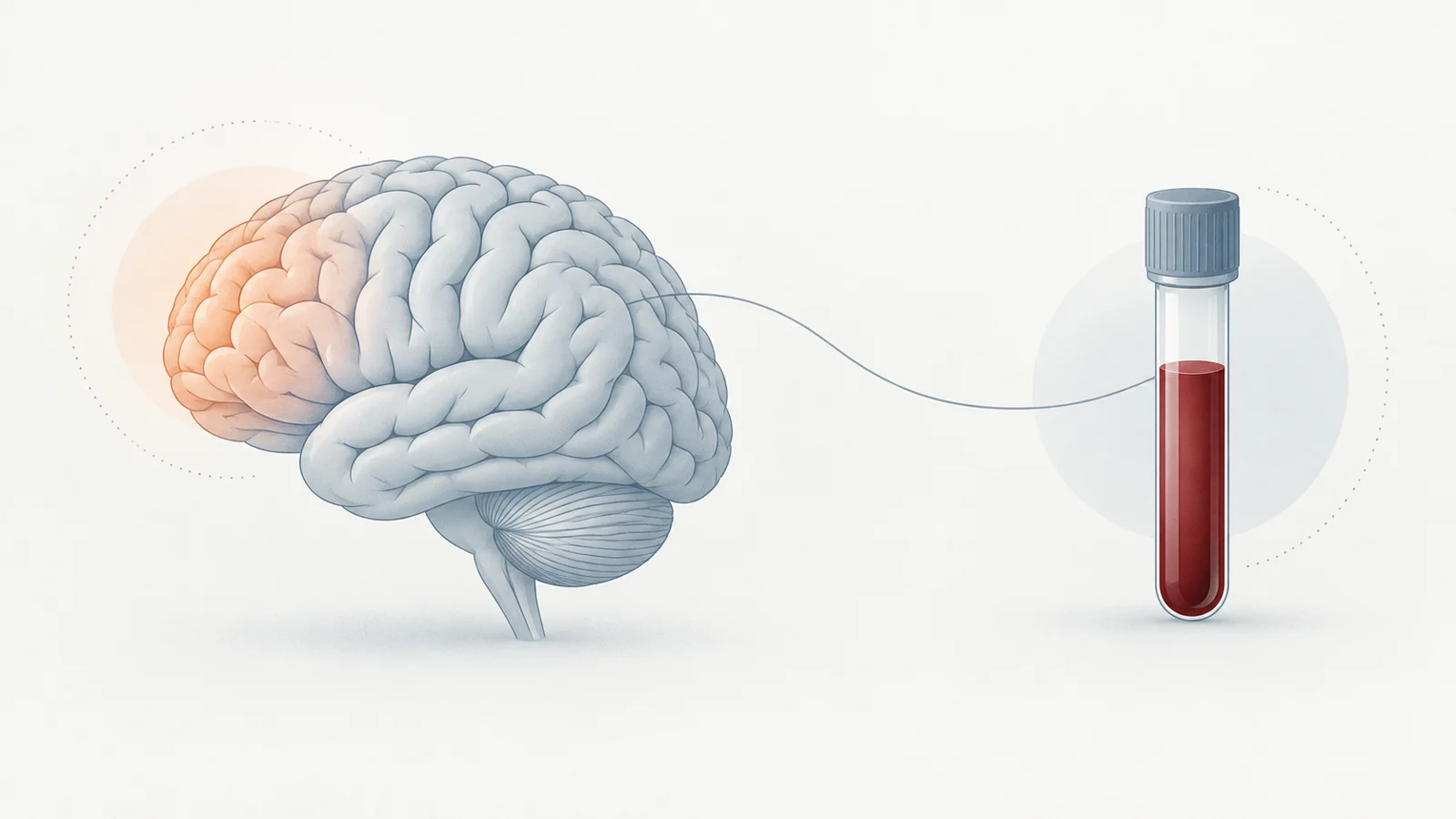
A study published on March 13, 2026, examined that question in active-duty Canadian Special Operations Forces (SOF) personnel. Brain scans showed higher uptake of a tau-targeting tracer in the exposed group’s frontal cortex, and blood tests found differences in markers associated with neural injury and astroglial activation. The results offer a snapshot of measurable differences in this small group. They do not establish that blast exposure caused them or show what will happen to participants over time.
A small comparison of active-duty personnel
The researchers compared 25 male SOF members with at least 16 years of work involving breaching, explosives or weapons use with 10 male military controls who had minimal blast exposure. Participants completed tau PET imaging, MRI, blood testing and clinical and cognitive assessments.
The groups also differed in their histories of head injury. A reported lifetime head injury was common among SOF participants, at 92%, compared with 30% of controls. Forty-four percent of the SOF group reported an injury involving loss of consciousness; no control participants did. This imbalance matters when interpreting group differences: the scan and blood patterns cannot be attributed to occupational blasts alone from this comparison.
The study was observational and cross-sectional: each person was assessed at one point in time. It could compare exposure histories and current measurements, but not trace when biological changes began.
The clearest scan difference was in the frontal cortex
The PET scans used [18F]flortaucipir, a tracer that binds to tau-related targets. Tau is a protein found in the brain; the scan measured where the tracer accumulated, not a confirmed disease diagnosis.
Average tracer uptake was higher in the exposed group’s frontal cortex than in controls, by 4.7%. That planned regional comparison remained statistically significant after the study’s correction for multiple comparisons. Uptake was also higher in the temporal cortex, by 5.3%, but that difference did not survive the correction. The frontal result is therefore the more robust of the two regional findings.
The researchers also compared individual scans with the control group’s measurements. Using their threshold for elevated uptake, they found clusters in 22 of the 25 exposed participants. Nearly half of those clusters were in frontal regions. This means the pattern appeared in many participants relative to this small control group; it does not mean that 88% had a diagnosed tau disorder.
More years of breaching tracked with higher uptake
Within the exposed group, participants reporting more years of breaching tended to have higher tracer uptake across several brain regions. Years of explosives work were also associated with higher uptake in some regions. For example, the relationship between breaching years and frontal uptake remained after adjustment for age.
A gradient across reported exposure histories makes the result relevant to the question of cumulative occupational risk. But years of work are not a direct measurement of the pressure wave each person experienced. Exposure histories were estimated retrospectively, and the study cannot separate blast effects from head injuries or other differences between participants. The associations describe how reported work history and scan measurements varied together, not proof of a causal pathway.
Blood measurements added related clues
Several plasma markers differed between groups. The exposed participants had higher levels of GFAP, a marker associated with astroglial activation; neurofilament light and UCH-L1, markers related to neural injury; and brain-derived tau, a measure associated with tau released from the central nervous system. Some phosphorylated tau measures were also higher, while others did not show significant group differences. The ratio of amyloid-beta 42 to amyloid-beta 40 was lower in the exposed group.
Selected markers also varied with regional PET uptake. GFAP and brain-derived tau, for example, were associated with uptake in several regions. These relationships provide a second kind of measurement that broadly corresponded with the scan findings in this sample. They do not validate a blood test for tau disease, establish a screening threshold or show that any individual’s result predicts future illness.
Scan uptake and reported difficulties were linked
Higher uptake in some brain regions was associated with more post-concussive symptoms, poorer sleep and lower duty-readiness scores. Within the SOF group, those associations were reported for several regions. Associations with PTSD and anxiety were weaker and did not remain significant after correction in that group.
These measures capture reported symptoms and functional assessments alongside brain imaging. Their association is a reason to study how the findings relate to service and wellbeing, but a one-time comparison cannot determine whether uptake preceded symptoms, followed them or reflected a shared factor. The results also do not provide an individual return-to-duty decision rule.
What earlier brain checks could change for blast-exposed personnel
If larger longitudinal studies confirm that these scan and blood patterns track meaningful changes over time, combining them could eventually help identify when brain-health assessments are useful for personnel with substantial exposure histories. That could support more targeted monitoring during service and help evaluate whether exposure-reduction measures are associated with healthier outcomes. The current study is an early step toward that possibility, not a ready-made screening or personnel-management tool.
Its small, largely white male sample limits how well the findings apply to women, more diverse military groups or civilians. Retrospective exposure estimates, unequal head-injury histories and the cross-sectional design complicate attribution. Flortaucipir can also bind outside its intended target, so uptake alone cannot establish chronic traumatic encephalopathy or predict future neurodegeneration. Following participants over time with more specific tracers and better exposure measurements would clarify whether the observed differences persist or progress.
Key Sources
- Primary source: Tau-PET imaging and blood biomarkers reveal early tauopathy in special operations forces exposed to repetitive blast.
DOI or Journal Page · Peer-Reviewed Paper · 2026-03-13
Keep exploring