
Research Signal – Source date 2026-01-20
An aging ovary can lose eggs and also become less able to support the eggs that remain. Those changes are related, but they are not identical: the follicle pool shrinks over time, while the quality of an individual oocyte—the cell that can mature into an egg—can decline as its surrounding environment changes.
A review published on January 20, 2026, brings these problems together through a metabolic account of ovarian aging. It describes how energy production, nutrient use, gene regulation and immune activity may influence one another inside the ovary. The proposed connections help explain why egg quality may suffer alongside falling egg numbers. They also point to possible intervention targets, although the review does not establish that any of them preserve fertility in people.
An egg depends on its follicle
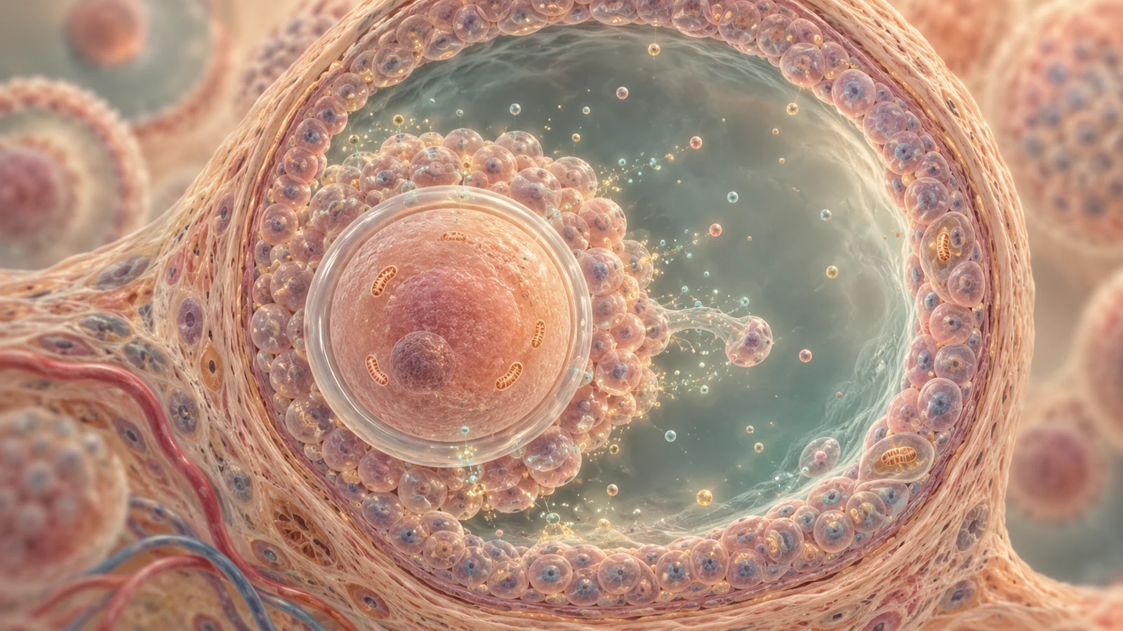
A follicle is a small working unit, with an oocyte at its center and supporting cells around it. Those surrounding cells help supply the oocyte with molecules it needs to maintain energy balance and prepare for development. The oocyte, in turn, sends signals that shape how the support cells use glucose and cholesterol.
This partnership matters because the oocyte does not handle every metabolic task on its own. Cumulus cells, which closely surround it, can provide substrates such as pyruvate, hypoxanthine and NADPH. The cells exchange small molecules through channels between them, and they also communicate through secreted signals and extracellular vesicles that carry regulatory molecules. These routes help coordinate energy use, stress responses and the conditions needed for the oocyte to mature.
The review describes evidence that this exchange can weaken with age-related dysfunction. When communication through gap junctions declines, or the signals carried between cells become disrupted, metabolite transfer may become less efficient. Supporting cells can also lose some of their ability to protect the oocyte from oxidative stress. The result can be a developmentally less capable oocyte even before the follicle itself is lost.
Follicles have substantial energy demands. Oocytes contain many mitochondria, and the surrounding cells multiply as a follicle grows. The ovary also uses energy to make steroid hormones. In the review’s account, these demands make the follicle particularly sensitive to problems in the systems that supply and allocate energy.
When mitochondria falter, glycolysis takes on more work
Mitochondria help cells make usable energy through oxidative phosphorylation. The review describes age-related damage to mitochondrial DNA, changes in the electron transport chain and failures in mitochondrial quality control. When mitochondrial energy production becomes less efficient, cells can face an energy shortage and produce more reactive oxygen species, chemically reactive molecules that can add to cellular stress.
One response is to increase glycolysis, a way of breaking down glucose that does not depend on the same mitochondrial process. Early on, this shift may help meet energy needs. But persistent or poorly regulated compensation can alter the follicle’s chemical environment. In the studies synthesized by the review, changes in glycolysis are linked to metabolite buildup, impaired signaling and reduced function in both oocytes and supporting granulosa cells.
The response may also change over time. The review describes a three-stage pattern in granulosa cells from patients with occult premature ovarian insufficiency: reduced glucose uptake, a compensatory rise in glycolysis, and eventual ATP depletion as mitochondrial compensation fails. This sequence is one model drawn from the evidence discussed in the paper; it should not be read as a universal timeline for every person with ovarian decline.
Metabolic coupling between the oocyte and its support cells adds another layer. For example, glycolytic activity in granulosa cells around ovulation has been associated with oocyte developmental competence. If the cells cannot provide the right balance of energy-related molecules, an increase in one pathway may not restore the coordinated support that a follicle needs. Metabolism here is not simply a matter of producing more energy; timing and exchange between cells also matter.
Metabolites can influence gene activity and inflammation
The review’s central proposal is that metabolism, epigenetic regulation and immune activity can reinforce one another. Epigenetic processes help regulate which genes are active without changing the underlying DNA sequence. Some enzymes involved in these processes depend on metabolites, so changes in cellular energy and nutrient availability can affect gene regulation.
NAD+ is one example. Cells use it in metabolic reactions, and it also serves as a substrate for sirtuins, enzymes involved in gene regulation, DNA repair and other processes. The review connects reduced NAD+ availability with weaker sirtuin activity and mitochondrial stress. In the proposed cycle, that stress can increase oxidative damage and disrupt the cell’s ability to manage energy and repair DNA.
Gene regulation may also shape inflammatory activity. The review discusses findings involving HDAC4, a regulator of gene activity. Its loss is linked in the reviewed studies to greater activity of an inflammatory and tissue-remodeling program associated with senescent cells. Senescent cells no longer divide normally and can release a mixture of inflammatory signals and enzymes known as the senescence-associated secretory phenotype, or SASP.
Those signals may help maintain a local inflammatory environment. The review also connects NAD+ depletion and altered glycolysis with SASP activity in granulosa cells. In this account, inflammation can worsen mitochondrial and metabolic stress, while stress-related changes can encourage further inflammatory signaling. The links come from different kinds of studies and do not show that every step occurs as one chain in every aging ovary. They offer a way to understand how several processes might compound one another.
Lipids and nutrients create other kinds of stress
Not all metabolic routes enter the proposed cycle through energy production. The review also describes lipid exposure around the ovary. After menopause, lower estrogen is associated with changes in peri-ovarian fat, including more pro-inflammatory macrophages and release of free fatty acids and inflammatory factors. These substances may reach ovarian tissue and contribute to lipid accumulation, mitochondrial dysfunction and local inflammation. This is a route from the surrounding tissue into the follicle’s environment, rather than simply a failure of the oocyte’s own energy machinery.
A separate lipid-related process is ferroptosis, a form of cell death driven by iron-dependent lipid damage. The review discusses findings linking increased ACSL4 expression and reduced GPX4 activity with lipid peroxidation in oocytes. It also describes experiments in which blocking ferroptosis-related processes improved measures of oocyte or ovarian function in models. These findings make lipid damage a potential point of study, but they do not demonstrate a fertility-preserving treatment for people.
Nutrient metabolism raises different concerns. In aged oocytes, altered glutamine use may replenish components of the energy-producing cycle. Changes in serine, glycine and one-carbon metabolism can affect the availability of methyl donors used in DNA and RNA regulation, as well as precursors for antioxidants. The review also notes that oocytes depend on long-lived proteins, leaving them vulnerable when protein maintenance and clearance systems are impaired. These pathways connect nutrient handling to gene regulation, oxidative protection and the stability of cell structures, without making them interchangeable with lipid injury or mitochondrial failure.
Intervention ideas remain at different stages
The review gathers several possible intervention points. Some aim to improve mitochondrial quality control, including processes that remove or replace damaged mitochondria. Others target oxidative stress, excessive mitochondrial fission, ferroptosis, senescent cells or the local tissue environment. The range reflects the review’s view that ovarian aging may involve several connected mechanisms, rather than one switch that can simply be turned back on.
The evidence behind these ideas is varied. The review discusses mechanistic studies, observations in human tissue or clinical populations, and interventions tested in animal or other experimental models. For example, some model studies report improved ovarian measures after mitochondrial or ferroptosis-related interventions. Such results can help identify pathways worth investigating, but they are not equivalent to evidence that a treatment preserves egg quality, fertility or hormone function in people.
The review also proposes future work on biomarkers and personalized approaches. A useful marker might help distinguish which processes are most active in a particular person’s ovarian decline. But a marker-based care pathway would first need validation: researchers would need to show that the marker identifies a meaningful biological pattern, that a matched intervention can safely reach its target, and that treatment improves outcomes people value. The article presents this as a direction for research, not an established clinical option.
What this could change for fertility care
If researchers can validate which metabolic and inflammatory processes matter most for different patterns of ovarian decline, fertility care could eventually match protective strategies to the biology most relevant to an individual. That might help avoid treating ovarian aging as one uniform process and focus attention on interventions with a plausible target in a particular case. Reaching that point depends on reliable markers, safe delivery and human studies showing meaningful benefits for ovarian function or fertility; the review describes a rationale for pursuing that work, not a proven way to preserve eggs.
Key Sources
- Primary source: Metabolic, epigenetic, and immune crosstalk in ovarian aging.
DOI or Journal Page · Review or Meta-Analysis · 2026-01-20
Keep exploring