
Research Signal – Source date 2025-08-26
A protein from the brain’s support cells
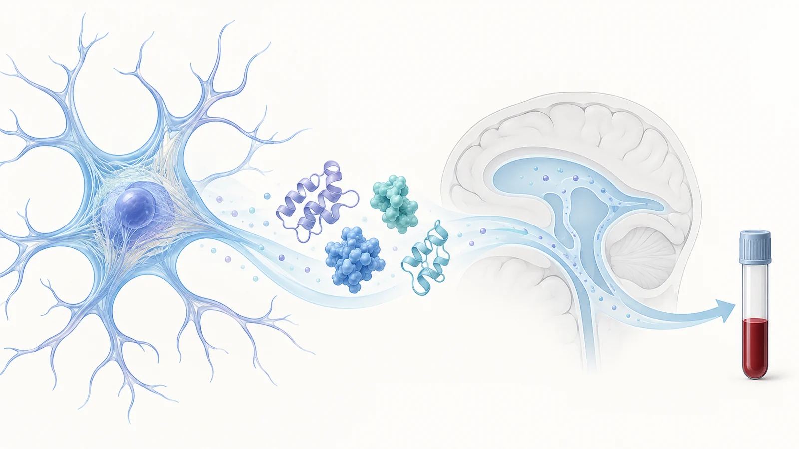
Glial fibrillary acidic protein, or GFAP, is a structural protein in astrocytes, cells that support the brain’s nerve cells. When the brain responds to injury or disease, astrocytes can become reactive and produce more GFAP. Some of that protein may leave the brain and reach the blood, where a test can measure it.
A review published on August 26, 2025, connects this biology with blood testing in traumatic brain injury (TBI) and Alzheimer’s disease (AD). Its central interpretive question is practical: when GFAP is elevated in either setting, what does the result say about the brain?
The answer depends on more than how much GFAP astrocytes produce. The protein’s physical form can affect what a test recognizes, and its route out of the brain can affect how much reaches the bloodstream.
A changing filament can become a different test target
Inside astrocytes, GFAP molecules assemble into filaments that help provide structural support. The protein’s units join in stages, building larger structures that can also be taken apart and recycled. This is a dynamic arrangement, not a fixed scaffold.
Chemical modifications help regulate that assembly. The review describes phosphorylation, in which phosphate groups are added to GFAP, as one process that can loosen filaments into soluble forms; removing those groups can support reassembly. Other changes can affect the protein’s stability, solubility, or susceptibility to cleavage into smaller fragments.
These changes matter at the test bench. Blood assays use antibodies that bind particular parts of a protein. A modification near an antibody’s target can make that region harder to recognize; cleavage can leave fragments that the usual assay does not detect. Some altered forms may also aggregate or become less soluble. As a result, a measured value reflects the forms an assay can capture, not necessarily every form of GFAP present.
The route to blood is part of the measurement
After GFAP is released from astrocytes into the space around brain cells, the review describes it moving through pathways around brain arteries, into areas where cerebrospinal and interstitial fluids exchange, and then along routes around veins and through lymphatic circulation toward the systemic blood.
That journey makes transport part of the biology behind a blood result. In AD, the review points to reported changes that may impede movement out of the brain, including altered aquaporin-4 distribution, weaker arterial pulsations, amyloid accumulation in glymphatic spaces, and sleep apnea. These are proposed obstacles to GFAP’s exit, not simply changes in its production.
After TBI, the review describes a different context for disrupted clearance. Raised pressure and changes in meningeal lymphatic vessels may reduce the movement of fluid and solutes out of the brain. The cited animal work includes a substantial reduction in lymphatic drainage after injury, with abnormalities described as persisting for one to two months. Those findings concern drainage and tissue changes in animal models; they do not provide a direct conversion from an individual patient’s blood value to the amount of brain injury.
In head injury, the question is about acute assessment
For TBI, the review discusses GFAP alongside UCH-L1, another blood marker, in studies of injury assessment. A multicenter observational study summarized by the authors reported that the paired markers had 0.976 sensitivity and 0.996 negative predictive value for identifying neuronal injury. In that study, fewer than one percent of cases with a negative blood test had a positive CT scan.
The reported use is as an aid in decisions about brain imaging, and the findings belong to the study populations and test context the review describes. The review also notes that both markers were detectable within hours after injury and could remain detectable for days. Together, these observations address an acute clinical question: whether blood markers may help clinicians assess injury and decide whether imaging is needed.
They do not establish a GFAP-only Alzheimer’s test. The review describes an FDA-approved i-STAT test for TBI that measures GFAP and UCH-L1 in blood collected within 24 hours of injury. That approval concerns the paired TBI test; it does not establish an approved GFAP-only diagnostic use for AD.
In Alzheimer’s studies, the question is about amyloid and cognitive stage
In the AD studies summarized by the review, blood GFAP is considered in relation to amyloid status and cognitive groups. One study reported higher plasma GFAP among amyloid-positive than amyloid-negative participants. In the TRIAD study, plasma levels differed across groups including cognitively unimpaired people with amyloid, people with mild cognitive impairment, people with AD, and cognitively normal, amyloid-negative participants. The review reports that plasma GFAP distinguished amyloid-positive from amyloid-negative groups better than cerebrospinal-fluid GFAP in that study.
Other findings described in the review link higher GFAP with brain imaging measures and with later cognitive outcomes. For example, a cited study associated higher blood GFAP with hippocampal volume loss and a greater risk of cognitive deficits in older adults. These are relationships observed across study groups and follow-up periods. They do not yield a GFAP-only diagnosis for one person or a reliable timetable for when an individual with mild cognitive impairment will develop dementia.
The purpose differs from the acute TBI question. In these AD studies, the focus is on how GFAP levels relate to amyloid and cognitive status across groups, rather than whether a paired marker result can inform an imaging decision after a recent injury.
The evidence depends on context
The authors identify limits in the evidence base: their review search used PubMed alone, and GFAP can rise in neurological conditions beyond AD, including other forms of dementia and injury. They call for clinical studies across different settings and age groups, and for better understanding of how GFAP moves from brain to blood.
A blood result also cannot, on the evidence summarized here, separate how much GFAP was produced from how its molecular form affected assay recognition or how transport affected its arrival in blood. Those influences are relevant to interpretation, but the review does not establish one threshold that applies across TBI and AD or across the varied studies it discusses.
How a dependable GFAP blood signal could guide care
If context-specific blood tests can be validated alongside a clearer understanding of GFAP’s molecular forms and movement into circulation, clinicians could gain a less invasive way to assess brain-related changes. In acute head injury, paired markers might help inform imaging decisions; in Alzheimer’s research and care, GFAP could contribute to understanding amyloid status and cognitive change. Those uses depend on studies establishing how reliably each test works for its intended population and question.
Key Sources
- Primary source: Deciphering the Structural Biology of GFAP: Connotations of Its Potency in Presaging the Diagnosis for Traumatic Brain Injury and AD.
DOI or Journal Page · Review or Meta-Analysis · 2025-08-26
Keep exploring