
Research Signal – Source date 2024-02-08
Most joint imaging shows the condition of tissue: narrowed spaces, damaged cartilage, swelling or other structural changes. A different kind of scan would ask a more specific question: are cells inside or around the joint entering a state that may help sustain inflammation?
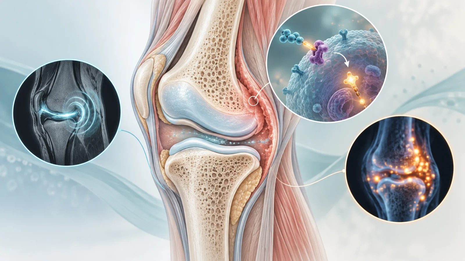
That question matters because senescent cells stop dividing but remain biologically active. The February 8, 2024 review Musculoskeletal imaging of senescence describes experimental MRI and PET approaches intended to detect an enzyme activity associated with these cells. The work is aimed at making senescence visible inside musculoskeletal tissues, but the reported evidence remains preclinical and investigational rather than a validated test for people with arthritis.
Why these cells matter inside a joint
Senescence is a response to aging or stress in which a cell enters a lasting growth arrest. The affected cell does not simply become inert. It can continue releasing inflammatory substances known collectively as the senescence-associated secretory phenotype, or SASP.
The review places this biology in cartilage, synovium and bone marrow around affected joints. A temporary senescence response can support healing and other protective processes. When the state persists, however, inflammatory secretion may help maintain tissue damage. In osteoarthritis, the review describes effects on the chondroid matrix and the recruitment of macrophages. In rheumatoid arthritis, it links SASP with ongoing joint inflammation.
Those observations do not make senescent cells the sole cause of either disease. They identify a cellular process that may contribute to the local environment and raise a practical imaging problem: tissue assays can reveal senescence after material is removed, but they are not a convenient way to repeatedly examine the same living joint during disease or treatment.
A scan would read an enzyme, not a cell count
The experimental probes discussed in the review target senescence-associated beta-galactosidase, usually abbreviated SA-β-Gal. This is an enzyme activity commonly used to identify senescent cells in laboratory samples. The imaging approaches use it as a biological trigger for a detectable change.
For MRI, the review describes a gadolinium-containing probe designed to be cleaved by SA-β-Gal. Once activated in the presence of the enzyme, the probe produces stronger signal on T1-weighted MR images. The intended result is not a photograph of individual cells. It is an enzyme-sensitive change in signal that can be compared with the tissue location where senescent cells are suspected.
The PET approach uses a different physical readout. A radioactive substrate is designed to be cleaved by SA-β-Gal and retained inside the lysosomes of senescent cells. Material that is not cleaved is eliminated through the kidneys, while retained tracer can be visualized with PET and related imaging. In principle, this could provide a way to detect and quantify tracer accumulation in a joint region, but the signal remains a proxy for the targeted enzyme activity rather than a validated absolute count of senescent cells.
Traditional MRI is already useful for evaluating musculoskeletal pathology, yet the review explains that conventional techniques do not specifically identify senescence. Histology, fluorescence microscopy, immunohistochemistry and flow cytometry can detect SA-β-Gal and related markers in tissue samples, but those methods do not provide repeated, non-invasive measurements in the same living subject.
What the experimental images have shown
The review reports that cartilage regions showing enhancement with the SA-β-Gal-sensitive MRI probe corresponded with histopathological evidence of senescent cells. Areas of intact cartilage did not show the same probe retention or MRI signal in the described observations. The agreement between imaging and tissue examination supports the idea that the probe was responding in locations enriched for the targeted cellular process.
The correspondence links the MRI signal with a tissue-based reference, supporting further investigation of the probe. It does not establish that the scan can reliably detect senescence in people with naturally occurring osteoarthritis or rheumatoid arthritis.
The PET experiments used more deliberately constructed animal models. In mice, researchers induced senescence in joints with doxorubicin and then looked for retention of SA-β-Gal-targeted radiotracers. In pigs, doxorubicin-treated senescent cells were implanted into knee joints. PET/MR imaging showed tracer retention in the joints containing the induced or implanted senescent material, while untreated joints and skeletal muscle used as a reference did not show the same retention pattern.
These controls make the model-level interpretation more persuasive: under the conditions described, the tracer distinguished joints prepared to contain SA-β-Gal-expressing senescent cells from comparison tissues. They do not establish performance in naturally occurring osteoarthritis or rheumatoid arthritis, where disease biology, tissue composition and the distribution of senescent cells may differ from an experimentally prepared joint.
The gap between an experimental signal and patient care
At the review’s February 2024 perspective, no diagnostic test had been clinically established to detect and quantify cellular senescence in patients’ articular joints. The authors describe groups working toward investigational applications for both MRI and PET probes, including the regulatory and approval steps needed for clinical translation. That work signals an effort to move beyond laboratory and animal studies; it does not mean that these probes were approved or routinely available.
Three claims therefore need to remain separate. The first is that an imaging signal can appear in an experimental model and correspond with tissue evidence. The second is that a probe can become a reliable diagnostic biomarker in people with arthritis. The third is that using the biomarker to choose or adjust treatment improves pain, mobility, cartilage preservation or another meaningful outcome. The evidence summarized here supports the first claim, while the latter two remain future clinical questions.
The proposed treatment connection involves senolytics, therapies intended to clear senescent cells. The review presents imaging as a possible way to identify patients who might benefit and to follow changes during treatment. It also emphasizes that senolytic safety and efficacy in musculoskeletal disease require further investigation. Imaging cannot resolve those therapeutic questions by itself; it could become useful only if the probes prove clinically valid and the treatments prove safe and beneficial.
What targeted scans could change for arthritis care
For someone with arthritis, the practical promise is more specific than simply seeing a damaged joint. A validated scan might help show whether the targeted senescence-related process is present in a particular joint and allow clinicians to follow its behavior over time. That could eventually support more informed selection or adjustment of treatments intended to clear senescent cells.
This benefit is conditional on several links holding together: the enzyme-sensitive MRI or PET signal must reliably reflect the relevant biology in patients, the test must be usable repeatedly, and senolytic therapies must demonstrate meaningful benefits without unacceptable risks. From the review’s February 2024 perspective, the field had developed a plausible route from tissue detection to repeated imaging, but it had not yet completed the journey to routine arthritis care.
Key Sources
- Primary source: Musculoskeletal imaging of senescence.
DOI or Journal Page · Review or Meta-Analysis · 2024-02-08
Keep exploring