
Research Signal – Source date 2024-01-17
A basic question sits beneath many discussions of Alzheimer’s disease. If oxidative damage and senescent cells contribute to the disease, why would antioxidants or drugs that remove senescent cells not simply protect memory?
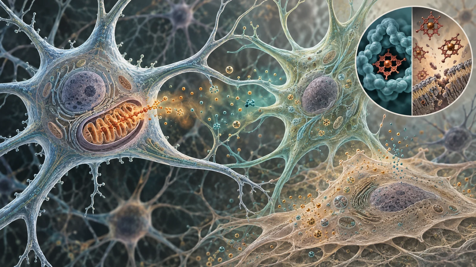
The answer is that these processes are part of a tightly connected biological system, not isolated switches. Oxidation is necessary for life, senescent cells are not identical to dead cells, and findings from a mouse brain do not automatically predict a meaningful benefit for a person. A review published on January 17, 2024, brings these complications together by examining redox changes, cellular senescence, amyloid, tau, mitochondria and heme metabolism in Alzheimer’s disease.
When useful chemistry becomes cellular damage
Redox chemistry describes reactions in which molecules gain or lose electrons. These reactions help cells produce energy, communicate, defend themselves and repair tissue. The problem begins when the balance between reactive molecules and the systems that control them shifts too far toward damage.
Reactive oxygen species, often shortened to ROS, are chemically active molecules produced during normal cellular activity. In excess, they can damage proteins, fats and DNA. The review describes evidence of increased oxidative-stress markers in brain tissue and biological fluids from people with Alzheimer’s disease or mild cognitive impairment. It also discusses reductions in antioxidant enzymes, including components of the glutathione and thioredoxin systems, in parts of the nervous system.
This does not mean that oxidation is itself an abnormal process. It means that the protective and restorative systems may no longer keep pace with the damaging reactions. Once proteins, membranes or genetic material are impaired, neurons can function less effectively and may eventually die. The same chemical pressure can also push living cells into a persistent stress state known as cellular senescence.
Senescence is more than a cell stopping
Cellular senescence is often introduced as a state in which a cell permanently stops dividing. That description is useful for many dividing cells, but it is incomplete for the brain. Neurons are already post-mitotic, meaning they normally do not divide. In Alzheimer’s disease, the important question is not simply whether a neuron has newly stopped dividing. It is whether the cell has developed senescence-associated features that alter how it functions and communicates.
The review describes increased markers associated with senescence in neurons, astrocytes and microglia, including senescence-associated beta-galactosidase, p53, DNA or telomere damage and the senescence-associated secretory phenotype. The last term refers to the release of signaling molecules such as inflammatory cytokines, chemokines and metalloproteinases.
Those secretions can change the environment around the cell. They may intensify inflammation, disrupt synapses and place additional stress on nearby neurons. A senescent cell is therefore not simply a cell that has disappeared from the tissue. It may remain active while contributing to a more damaging local environment. Excess reactive oxygen molecules can promote this state, while senescence-associated mitochondrial problems can generate still more reactive molecules.
The review presents this relationship as mutually reinforcing, but it does not establish one universal starting point in people with Alzheimer’s disease. Oxidative stress, senescence, inflammation and neuronal dysfunction may influence one another in different ways across cells and stages of disease.
Amyloid, tau and mitochondria in the same conversation
Amyloid and tau fit into this biology as interacting contributors rather than as explanations that stand apart from cellular chemistry. The review discusses evidence that abnormal amyloid-beta and tau are associated with redox imbalance, mitochondrial dysfunction, endoplasmic-reticulum stress, calcium disturbance and inflammatory responses.
In experimental systems, amyloid exposure can increase reactive oxygen production and senescence-related markers. The review also describes findings in which oxidative stress promotes the formation of hyperphosphorylated tau through changes in enzymes that regulate tau. Conversely, pathological tau has been linked to increased reactive oxygen molecules and greater sensitivity to stress-induced neuronal death in a transgenic model.
Mitochondria add another feedback point. They are important sources of reactive oxygen molecules because of their role in energy production. Damage to mitochondria can reduce membrane potential and respiratory capacity, making it harder for the cell to maintain normal function. The review describes long-term neuronal cultures in which changes in mitochondrial membrane potential accompanied increased ROS and a larger proportion of cells with senescence-associated features.
Human observations point in a similar direction without resolving the sequence of events. Studies discussed in the review found more oxidized DNA bases, including damage in mitochondrial DNA, in Alzheimer’s samples than in control samples. These findings show that damaged cellular chemistry is associated with the disease. They do not by themselves show whether oxidative stress starts the process, follows other pathology, or varies by cell type and disease context.
Heme shows why balance matters
Heme makes the treatment problem more concrete. It is an iron-containing molecule used by proteins involved in oxygen transport, mitochondrial electron transfer, storage and cellular signaling. Bound within the right proteins and compartments, heme supports essential functions.
Free heme and excess iron are different. When heme is released from its normal setting, or when heme availability and breakdown become disturbed, iron-containing chemistry can promote oxidative stress and lipid peroxidation. The resulting damage can affect membranes and contribute to cell death. At the same time, too little usable heme can impair mitochondrial activity and other necessary processes.
The review discusses evidence that altered heme handling affects heme proteins, iron regulation and mitochondrial function in Alzheimer’s disease. It also describes interactions between heme and amyloid-beta or tau that are associated with plaques and neurofibrillary tangles. In one line of experimental work, a heme–amyloid complex generated oxidizing activity and reduced neurotransmitters such as serotonin. Other findings linked heme–amyloid interactions to mitochondrial-dependent neuronal death.
This is why “remove the damaging molecule” is not a complete treatment strategy. A useful intervention would need to limit harmful free heme reactions while preserving the heme-dependent chemistry cells need. The same principle applies to reactive oxygen molecules more broadly: reducing every oxidation reaction would not be compatible with normal life.
What different experiments can and cannot show
The review combines several kinds of evidence, and each answers a different question. Human brain samples, cerebrospinal fluid, plasma and urine can show that oxidative-stress markers or antioxidant systems differ in people with Alzheimer’s disease or mild cognitive impairment. Patient-derived cells can reveal disease-associated cellular behavior under controlled conditions. These observations help identify patterns linked to the disease, but they do not demonstrate that changing one measured pathway will improve memory.
Cultured cells and animal models allow researchers to manipulate amyloid exposure, oxidative stress or senescence and then observe molecular or cellular responses. For example, the review describes amyloid exposure increasing ROS and senescence-related features in cultured cells, and Alzheimer’s mouse models showing abnormal mitochondria, elevated ROS or markers associated with cellular senescence.
Those experiments can provide evidence for mechanism. They can show that a proposed pathway is biologically possible and that an intervention changes a measurable outcome under defined conditions. They cannot settle every question about human disease. A change in a cell marker, a mouse brain measurement or a mouse cognitive task is not the same as a sustained improvement in a patient’s memory, independence or everyday function.
Keeping these study designs separate also prevents a common shortcut. A human association can be treated as proof of causation, while a successful manipulation in a model can be treated as proof of clinical effectiveness. The review supports neither conclusion on its own.
Treatments target different parts of the problem
Antioxidants and senolytics aim at different points in the biology. Antioxidant approaches are intended to reduce oxidative injury or strengthen defenses against reactive molecules. Senolytics are intended to remove senescent cells and therefore reduce the harmful signals those cells may release.
The review describes mixed findings for antioxidant interventions involving vitamin E, vitamin C and selenium. Some studies associated antioxidant combinations with lower Alzheimer’s prevalence or slower functional decline, while other findings showed modest effects or no clear association. Selenium-related results included changes in measures such as glutathione peroxidase activity and cognitive performance, but the longer-term clinical meaning remained uncertain.
These outcomes should not be compressed into one label such as “worked” or “failed.” A biochemical marker, a cognitive measure and a functional outcome describe different levels of benefit. An intervention might alter an antioxidant enzyme without preserving daily abilities, or produce a modest change in function without correcting every underlying marker.
Senolytic treatment has a different evidence profile. The review discusses dasatinib and quercetin, a combination studied in Alzheimer’s mouse models. In those animals, the treatment reduced amyloid-beta 40 and 42 in the hippocampus and entorhinal cortex, eased plaque-associated inflammation and improved reported cognitive deficits. These are mouse findings, not evidence that the same benefits occur in people with Alzheimer’s disease.
The review also described human trial activity involving senolytics, including an Alzheimer’s trial identified as recently completed at the time of publication. Completion of a trial does not establish its efficacy, adverse-event profile or long-term outcomes. The review specifically notes that the short- and long-term safety of senolytic administration in humans remained unresolved.
Heme-directed ideas carry their own tradeoffs. The review discusses apoNeuroglobin as a way of sequestering heme from a heme–amyloid complex in experimental work, while noting that this approach had not been tested clinically. It also discusses methylene blue, including mixed clinical-trial findings at different doses and reported central-nervous-system and other adverse effects. These examples show why a pathway can be biologically attractive without yet providing a straightforward treatment for patients.
The practical question is therefore not whether oxidative stress or senescence is “real.” The more difficult question is whether an intervention can change the harmful part of that biology while preserving necessary cellular functions, and whether that change produces a meaningful benefit for people.
How this could expand Alzheimer’s treatment
A better understanding of redox imbalance, senescence-associated signaling and heme metabolism could eventually add treatment options that protect neuronal function by interrupting damaging processes before they erode cognition and everyday abilities. The potential benefit is conditional: selective interventions would need to preserve useful chemistry, act on the relevant cells and demonstrate meaningful patient benefit with acceptable adverse effects.
If those hurdles are solved, Alzheimer’s treatment could expand beyond approaches aimed at a single visible feature of the disease. It could include carefully targeted ways to reduce cellular damage and inflammatory signaling while keeping neurons and their supporting cells functional. That possibility would not come from the biology alone; it would depend on showing that the right intervention helps people retain cognitive and everyday abilities.
Key Sources
- Primary source: Redox changes and cellular senescence in Alzheimer's disease.
DOI or Journal Page · Review or Meta-Analysis · 2024-01-17
Keep exploring