
Research Signal – Source date 2022-11-07
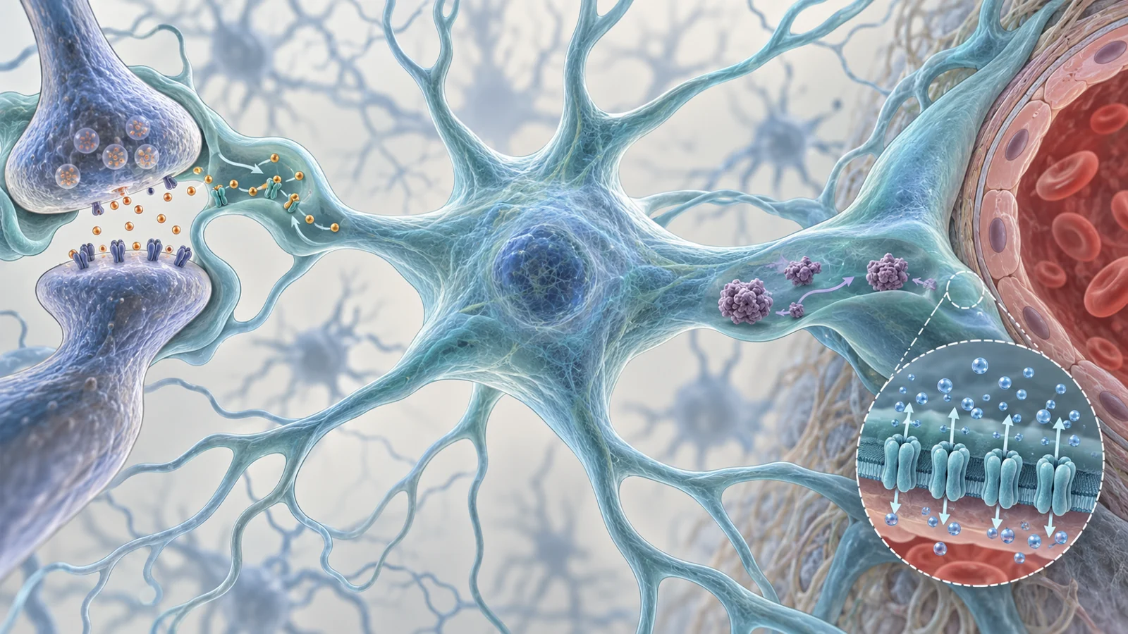
Alzheimer’s disease is usually discussed in terms of neurons, amyloid-beta and tau. But neurons do not operate alone. Astrocytes are support cells woven through the brain’s communication networks. They help supply metabolic resources, regulate ions and neurotransmitters around synapses, release signaling molecules and support the blood–brain barrier, the neurovascular unit and the glymphatic system involved in clearing substances from the central nervous system.
That makes them attractive therapeutic targets for a practical reason: changing the environment around neurons could influence how well those neurons communicate and cope with stress. It also creates a complication. Astrocytes can protect neurons in one setting and contribute to harmful inflammation, oxidative stress or disrupted signaling in another. The relevant question is therefore not simply whether astrocytes are “good” or “bad,” but which function is changing, where it is changing and whether an intervention can reach it safely.
A review published on November 7, 2022, examined these possibilities across laboratory cell systems, animal models, human observations and selected clinical findings. Its discussion suggests several routes for investigation, while keeping a central boundary in view: changing an astrocyte process in a model is not the same as preserving memory in people.
The support cells around neurons are active participants
One of the clearest ways to understand the therapeutic idea is to think of astrocytes as regulators of the space in which neurons work. They help control the concentration of chemical messengers outside cells, provide metabolic support and influence synaptic activity. They also communicate with microglia, the immune-related cells that respond to changes in the brain, and participate in processes connected to blood flow and waste clearance.
In Alzheimer’s disease, amyloid-beta can alter astrocyte behavior. The review describes changes involving calcium signaling, gliotransmission and inflammatory activity through pathways including RAGE and NF-κB. These changes can affect the balance between support and stress. Astrocytes may still perform protective tasks, but their responses can also add to inflammation or disturb neuronal communication, depending on disease stage and the surrounding cellular environment.
This is why an astrocyte treatment would need a precise aim. It might try to improve glutamate removal, alter lipid handling, reduce a harmful inflammatory response, support clearance or correct a calcium-related signal. Those are different biological jobs, with different delivery requirements and different standards for showing that a treatment helps.
When glutamate lingers, signaling can become damaging
Glutamate is a chemical messenger that helps neurons communicate. Its concentration outside cells has to be controlled: too little can interfere with signaling, while too much can overstimulate neurons. Astrocytes perform much of the cleanup. Under normal conditions, the review states that they remove about 90% of released glutamate in the central nervous system through transporters called EAAT1 and EAAT2, also known as GLAST-1 and GLT-1.
During Alzheimer’s progression, reduced astrocytic glutamate uptake can leave more of the messenger available to stimulate neuronal receptors. The resulting overactivation of NMDA receptors is part of a process described as excitotoxicity. In the review’s account, this involves impaired glutamate reuptake and increased glutamate release, with consequences that include synaptic loss and neuronal death.
The therapeutic logic is therefore more specific than “protect the brain.” Restoring a transporter could help keep chemical signaling within a useful range. In an Alzheimer’s mouse model, the review describes a pharmacological intervention that restored EAAT2 function. The treated mice showed improved cognitive function, fewer amyloid plaques and recovery of synaptic integrity in that experimental setting.
Those findings connect a cellular mechanism to several outcomes in the same animal model, but they do not show that increasing EAAT2 function preserves memory in people. They do, however, illustrate why astrocytes can become treatment targets before extensive neuronal loss: their role in regulating the surroundings of a synapse may be altered while the communication system remains partly modifiable.
Lipid handling connects astrocytes to amyloid
Astrocytes also influence the brain’s handling of fats. Cholesterol and other lipids are needed for cell membranes and signaling, but their transport and processing must remain coordinated. The review identifies astrocytes as the main producers of APOE in the brain. APOE is a protein involved in cholesterol transport, and its different forms are associated with different levels of Alzheimer’s risk.
This makes lipid metabolism relevant to amyloid-beta for two separate reasons. It can affect how material is taken into an astrocyte, and it can affect what happens after that material enters the cell. Uptake is not the same as degradation. A cell may internalize amyloid without breaking it down effectively, so a useful intervention would need to influence the relevant downstream processing as well.
The review describes an experimental example involving gemfibrozil, a drug used for dyslipidemia, combined with retinoic acid. In mouse-derived astrocyte systems, the combination enhanced amyloid-beta uptake through a PPARα-dependent route involving the low-density lipoprotein receptor. The review also links the treatment to TFEB, a transcription factor associated with autophagy and lysosomal function, and describes potential amyloid degradation and reduced plaque burden in the hippocampus of a 5xFAD mouse model.
This is a useful example of how a treatment hypothesis can be built from cell behavior to an animal outcome. It is not evidence that the combination is an Alzheimer’s treatment for patients. The findings concern experimental uptake, cellular processing and plaque-related outcomes, not a demonstrated improvement in human cognition, tolerability or safe dosing.
Location can decide whether a target works
A biological target is not just a molecule or pathway. It is also a question of location. The review presents two examples in which the place and route of an intervention matter, but they involve different mechanisms.
In one Alzheimer’s mouse model, astrocyte-produced interleukin-3, or IL-3, helped direct microglia to cluster around amyloid-beta plaques. The reported result included greater plaque clearance and improved memory in the model. Yet peripheral IL-3 injection did not change the pathology described in the review. The supplied account does not establish why the peripheral injection lacked an effect. It does show that producing or signaling a molecule within a particular brain-cell interaction cannot automatically be treated as equivalent to delivering the same molecule from outside the brain.
The second example concerns AQP4, a water-channel protein associated with astrocytes and the glymphatic system. Astrocytes have extensions called endfeet that are positioned alongside blood vessels. The review emphasizes that AQP4 would need to be targeted at these vessel-facing endfeet for a proposed clearance intervention to make biological sense. Increasing AQP4 elsewhere may not produce the same effect.
These examples point to two different spatial constraints. IL-3 involves the route and setting of communication between astrocytes and microglia. AQP4 involves the cellular subregion where astrocyte structure meets the vascular environment. In both cases, a promising molecular name is not enough. An intervention must reach the right cellular context and alter the function that matters there.
The hardest step is moving from mechanism to patient benefit
The review’s discussion of antioxidants shows why biological activity and clinical benefit have to be kept separate. In laboratory experiments, vitamin E improved astrocyte survival after glutamate-mediated toxicity in cultured cells. Other antioxidant systems also showed protective effects in preclinical Alzheimer’s models. But clinical trials of alpha-tocopherol, a form of vitamin E, reported limited benefits.
These findings answer different questions. A cell experiment can show that an intervention changes survival under a defined stress. An animal model can show effects on cognition, plaques or synaptic structure under experimental conditions. A clinical trial asks whether a treatment helps people, at an appropriate dose and timing, with effects that matter to their health. Positive results at one level do not automatically carry across to the next.
The review proposes several possible explanations for disappointing antioxidant results, including dosage, the timing of intervention, the type of antioxidant and incomplete understanding of how antioxidant systems operate. These are proposed explanations, not established reasons that settle the clinical question.
The same caution applies to human astrocyte cultures. A result in cultured human cells can make a mechanism more relevant to human biology than a result in a mouse cell, but it still does not establish clinical efficacy, tolerability or a safe dose in people. The authors also warn that animal models may not fully mirror human Alzheimer’s disease. That limitation matters especially when an experimental intervention changes amyloid uptake, transporter expression or plaque burden without directly demonstrating preserved human memory.
The review therefore treats astrocyte-focused approaches as future therapeutic directions rather than established care. Possible targets include inflammatory signaling, oxidative stress, glutamate transport, APOE-related lipid metabolism, calcium regulation and glymphatic function. A combination of mechanisms, such as pairing control of an inflammatory pathway with reduction of oxidative stress, is presented as a proposal for investigation rather than a proven treatment strategy.
What astrocyte therapies could mean for preserving memory
The potential benefit is practical and conditional. If an intervention could safely reach the relevant astrocyte function, maintain useful neuronal signaling and improve the handling of damaging proteins, it might help people in early Alzheimer’s retain cognitive function before extensive neuronal loss. The route would not be to replace neurons directly, but to preserve some of the cellular support that neurons need to communicate and remain resilient.
That possibility depends on several problems being solved at once: selecting the right astrocyte function, reaching the right location, avoiding harmful effects elsewhere and demonstrating meaningful benefits in people. The review does not establish that any of the discussed interventions meets those conditions. It identifies why astrocytes deserve attention as therapeutic targets and why their complexity makes translation difficult.
The most credible future use would therefore be a carefully targeted way to support the brain’s own communication and clearance systems, with patient benefit established through human evidence rather than inferred from a repaired transporter, altered plaque burden or protected cell in an experimental model.
Key Sources
- Primary source: Astrocytes as a Therapeutic Target in Alzheimer's Disease-Comprehensive Review and Recent Developments.
DOI or Journal Page · Review or Meta-Analysis · 2022-11-07
Keep exploring