
Research Signal – Source date 2022-10-26
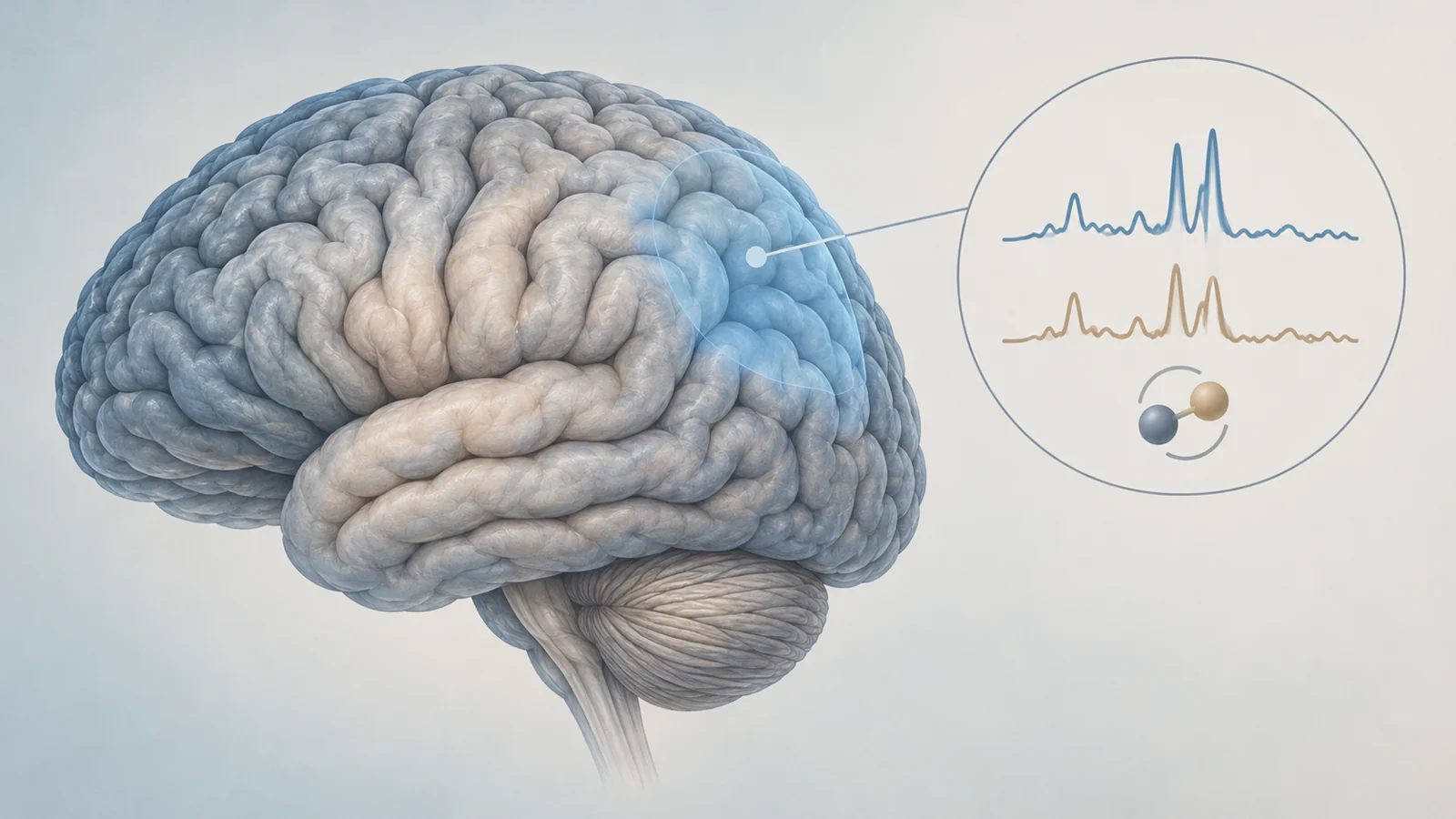
Perimenopause, the transition toward menopause, is associated with a higher risk of a major depressive episode. That raises a precise scientific question: does the transition also change chemical signals in brain regions involved in mood? A study published on October 26, 2022, tested that question in the left dorsolateral prefrontal cortex, a region toward the front and side of the brain.
In 38 healthy women, the scan comparison did not find a statistically significant difference between perimenopausal and reproductive-aged participants in either of the measured signals. The result applies to one brain region, two reference-based measurements and a group of women screened to be free of current or past psychiatric illness. It is not a finding that brain chemistry remains unchanged throughout perimenopause, nor does it explain why some women develop depression during the transition.
The scan asked a regional question
The dorsolateral prefrontal cortex is part of the outer frontal brain, behind the forehead and toward the side of the head. The researchers hypothesized that GABA+ and glutamate ratios in this left-sided region would be lower in perimenopausal women than in women of reproductive age.
Magnetic resonance spectroscopy, or MRS, is a non-invasive scanning method that estimates chemical signals within a selected volume of brain tissue. It differs from a structural scan that shows anatomy and from a functional scan that tracks changes in activity. Here, the researchers used a 3 Tesla scanner to measure GABA+ and glutamate in the selected left frontal region.
The plus sign in GABA+ matters. The reported signal includes GABA along with overlapping contributions from homocarnosine and macromolecules. The study therefore did not measure a pure GABA concentration. Glutamate was reported separately, but the signals were still expressed as ratios rather than as absolute amounts.
For both measurements, the reference was creatine plus phosphocreatine, written as Cr+PCr. Creatine was used here as a comparison signal within the spectroscopy measurement, not as a supplement or treatment. A ratio can show how one measured signal relates to the reference in the sampled tissue; it cannot by itself establish the absolute concentration of a neurotransmitter or the functional balance between excitatory and inhibitory communication.
Who was compared and under what conditions
The participants were 18 perimenopausal women and 20 women of reproductive age. A structured psychiatric interview screened participants for current or lifetime psychiatric illness. The researchers also excluded circumstances and medications that could affect brain GABA or glutamate function. Reported mood scores were within a range the authors considered normal fluctuations, and the perimenopausal participants reported little or no perimenopause-related symptom burden.
The comparison was designed to limit some variation in hormone timing. Reproductive-aged participants were scanned during the early follicular phase of the menstrual cycle. Early perimenopausal participants were scanned during an anovulatory cycle, while late perimenopausal participants had gone through three months without an ovulatory cycle. These choices made the sampling conditions more consistent within the study, but they cannot represent every hormonal state experienced during the transition.
The population also defines what the result can speak to. These were physically and mentally healthy women who were not experiencing clinically significant depressive symptoms. The findings therefore describe chemical measurements in a carefully screened group, not the brain chemistry of women currently experiencing depression.
What the numbers actually showed
The perimenopausal group had a mean left dorsolateral prefrontal GABA+ ratio of 0.10, compared with 0.11 in the reproductive-aged group. The comparison had a p value of 0.70. For glutamate, the corresponding means were 0.56 and 0.57, with a p value of 0.43.
Neither comparison reached the study’s threshold for statistical significance. An analysis that adjusted for age and the proportions of gray and white matter in the sampled region likewise found no significant effect of reproductive status on either ratio. The adjusted p values were 0.74 for GABA+ and 0.48 for glutamate, with small reported effect sizes.
These figures describe how close the group averages were in this sample. They do not establish that the two populations have identical brain chemistry, that every individual has the same measurements, or that perimenopause has no effect in other regions or under other hormonal conditions.
Why age and reproductive status cannot be separated here
The comparison involved different women at different points in life. The perimenopausal participants were 48.8 years old on average, while the reproductive-aged participants were 31.5 years old. Because age and reproductive status changed together between the groups, the design cannot cleanly determine whether a regional difference would reflect reproductive transition, aging, or a combination of both.
Statistical adjustment can account for measured differences in a model, but it does not turn a cross-sectional comparison into a study of the same person over time. A longitudinal design would be needed to follow individual brain measurements across the transition and more directly examine how they change.
The perimenopausal group was also small and unevenly divided between stages: 12 women were classified as early perimenopausal and six as late perimenopausal. That was not enough to make a meaningful comparison between those stages. The limited sample also leaves less ability to detect a modest difference, even though the group means were close. The authors argued that the observed differences were unlikely to represent a meaningful effect, but the study still cannot establish equivalence between the groups.
A contrast with the medial prefrontal cortex
The authors interpreted this result alongside earlier findings from their group involving the medial prefrontal cortex, a more centrally located frontal area behind the forehead. In that earlier work, they reported decreased GABA+ and glutamate ratios in perimenopausal women. Those earlier findings are background to the present paper’s interpretation; the current study did not directly measure both regions in the same participants as a head-to-head regional comparison.
The contrast led the authors to propose that different brain areas may have different sensitivity to the hormonal fluctuations associated with perimenopause. They also suggested that the medial region’s connections with subcortical areas involved in hormone signaling might help explain the discrepancy. That connectivity idea remains a hypothesis rather than a demonstrated mechanism in this study.
The measurements themselves also leave the biological source open. The researchers noted that GABA+ and glutamate signals may reflect neurotransmission, neuronal metabolism, or both. The scan therefore offers a regional chemical observation, not a direct readout of nerve-cell communication and not a measure that can explain depression in an individual woman.
What a regional brain map could reveal about perimenopausal depression
A reproducible pattern of regional chemistry could eventually help researchers identify which brain circuits participate in vulnerability to depression during the menopausal transition. That would make it easier to distinguish biological changes associated with the transition from changes specifically linked to illness, giving prevention research a more precise foundation for women who may be at risk.
That possibility depends on larger studies that separate age from reproductive status, sample the hormonal transition more fully and link regional measurements to later depression outcomes. The 2022 comparison offers one carefully bounded piece of that future picture: in healthy women, this particular left frontal region did not show a statistically significant group difference in the two reference-based signals that were measured.
Key Sources
- Primary source: A comparative magnetic resonance spectroscopy study of GABA+ and glutamate referenced to creatine and phosphocreatine in the left dorsolateral prefrontal cortex of perimenopausal women and women of reproductive age.
DOI or Journal Page · Peer-Reviewed Paper · 2022-10-26
Keep exploring