
Research Signal – Source date 2022-07-19
The inner surface of every blood vessel is lined by a thin living layer called the endothelium. It is more than a passive coating. Endothelial cells help keep blood fluid, regulate vessel width, control what passes from the bloodstream into tissues and help form new vessels when tissues need more oxygen or nutrients.
The review published on July 19, 2022, examines what happens when these cells become senescent. In this context, senescence means a lasting halt in cell division while the cell remains metabolically active and often becomes more secretory. A senescent endothelial cell is therefore not simply an old or dead cell. It can still alter the environment around it, including the behaviour of neighbouring cells.
That creates the central tension. How can a cell that has stopped multiplying continue to interfere with circulation and repair? The answer lies in the endothelium’s location and responsibilities. Its position at the boundary between blood and tissue exposes it to mechanical forces, circulating substances and signals from nearby cells. When those pressures activate damage responses, the resulting cell may stop dividing but continue sending disruptive signals.
Blood flow gives different endothelial cells different risks
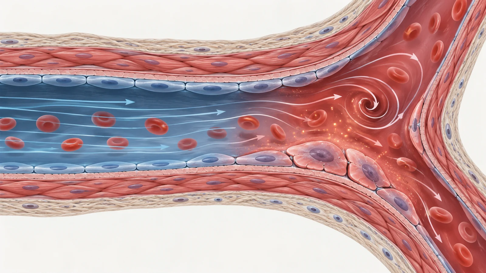
Endothelial cells do not experience the same physical environment throughout the vascular tree. Cells along relatively orderly, high laminar flow can receive signals associated with vascular protection. Cells at branches and curves, such as areas around the carotid sinus and aortic arch, experience low or oscillating shear stress. The review describes this disturbed flow as damaging and as capable of producing a phenotype resembling the inflammatory secretory behaviour associated with senescence.
Pressure and pulsatile forces add another source of strain, particularly in large arteries. At the same time, endothelial cells are continuously exposed to substances carried in blood, including oxygen, glucose, lipids, amino-acid metabolites, immune cells, reactive oxygen species and inflammatory cytokines. Some endothelial cells divide frequently in areas exposed to disturbed flow, creating repeated replicative stress. Others can remain in place for many years, accumulating damage through prolonged exposure to harmful conditions.
This means endothelial senescence is not explained by chronological age alone. Both frequently dividing cells and long-lived cells can reach a senescent state, but the route may differ. Damage to DNA, changes in energy-sensitive pathways and activation of tumour-suppressor responses can converge on permanent growth arrest and changes in cell shape and secretion.
Chromosome ends add an important detail. Telomeres help protect the ends of chromosomes, and their DNA sequence is vulnerable to oxidative damage. The review highlights evidence that oxidative damage can cause telomere uncapping, meaning loss of protective structure, even when telomeres are not unusually short. In older people and people with hypertension, telomere uncapping in endothelial cells has been associated with p53–p21-related senescence independently of telomere length. The implication is that measuring length alone may miss a relevant form of vascular damage.
A stopped cell can still change the vessel around it
Two endothelial jobs make the consequences easier to understand. One is signalling to the smooth muscle in the vessel wall so the vessel can widen or narrow. The other is controlling the barrier between blood and surrounding tissue.
For vessel relaxation, nitric oxide is especially important. Endothelial cells produce it and release it toward vascular smooth-muscle cells, where it promotes widening. Nitric oxide also helps limit the display of adhesion molecules, supports barrier function and discourages coagulation. The review describes evidence that nitric oxide can help prevent endothelial senescence, while senescent endothelial cells produce less of it and show reduced expression of the enzyme involved in its production.
This creates a potentially self-reinforcing problem. Less nitric oxide can weaken a protective signal, while senescent cells may further reduce nitric-oxide availability. The result is a plausible route from cellular dysfunction to impaired endothelium-dependent dilation, the ability of an artery to widen in response to signals from its lining. Yet the review is careful about the strength of that inference: endothelial senescence alone had not been shown unequivocally to be sufficient to disrupt this response.
The second job is selective permeability. Endothelial junctions regulate which molecules and cells move between the bloodstream and tissue. In cultured endothelial cells driven into senescence, junctional proteins including cadherin-5 and ZO-1 become disorganized, while occludin and claudin-5 are reduced. The consequence is not necessarily confined to the senescent cell itself. When senescent and non-senescent endothelial cells are grown together, the non-senescent cells can also develop dysfunctional tight junctions.
Inflammatory secretions from senescent cells may help explain this bystander effect. A more permeable endothelial layer can alter tissue boundaries, and the review connects endothelial senescence with impaired blood–brain barrier function in experimental settings. The broader point is that the secretory behaviour of a senescent cell can extend its influence beyond the cell that first sustained the damage.
Vessel growth can be impaired or become disordered
New vessel formation, or angiogenesis, is essential when exercise increases tissue demand or when injured tissue needs to restore its blood supply. Endothelial cells initiate and sustain this process through responses involving signals such as vascular endothelial growth factor, fibroblast growth factor and hypoxia-inducible factor 1-alpha. If senescence reduces the ability of endothelial cells to respond, tissues may have more difficulty building the vessels needed for recovery.
Evidence reviewed in mouse models supports that concern. Reducing endothelial senescence through genetic deletion of p21 or p53 improved proliferation and angiogenesis in some settings. Endothelial p53 deletion also improved angiogenesis and prevented pressure-overload cardiac fibrosis and diabetic cardiomyopathy in the reported models.
But senescence does not point in only one direction. In a mouse model of retinopathy discussed in the review, endothelial senescence promoted pathological angiogenesis, along with secretory factors associated with senescence. Senolytic treatment reduced disease severity by reducing this abnormal vessel growth while allowing functional vessels to regrow. Here, the problem was not simply too little angiogenesis. It was the wrong kind of vessel growth in the wrong tissue context.
That exception matters because it changes what a possible treatment would need to accomplish. A useful intervention might have to restrain pathological signals without blocking the vessel formation required for repair. The same broad process—new vessel growth—can be beneficial in one setting and damaging in another.
Experimental design changes what the evidence can show
Several lines of evidence connect endothelial senescence with vascular and metabolic dysfunction, but they do not all support the same level of causal inference.
Human observations can reveal associations. In healthy sedentary adults, higher endothelial senescence was associated with poorer endothelium-dependent dilation. Such findings are relevant to human physiology, but an association between markers and vascular function cannot by itself establish that senescent endothelial cells caused the impairment.
Animal experiments can manipulate the system more directly. In aged mice, the senolytic combination dasatinib and quercetin improved carotid artery dilation and reduced uncapped telomeres in aortic endothelial cells. The smooth-muscle response to nitric oxide did not change in that study, which supports an endothelial contribution to the observed vascular improvement. Even so, a whole-body senolytic intervention does not isolate endothelial cells as the only cells involved in the benefit.
Endothelial-specific genetic experiments provide a sharper test. In mice, endothelial-cell-specific changes involving NF-kappa-B, p53 or the telomere-capping protein TRF2 altered inflammatory signalling, insulin resistance and related metabolic outcomes. For example, deleting endothelial Trp53 reduced obesity, glucose intolerance and insulin resistance in one set of experiments, while increasing endothelial p53 worsened those phenotypes. Senescent endothelial cells also released factors that affected adipocytes and impaired insulin signalling in those experimental systems.
These results make endothelial senescence a plausible contributor to systemic metabolic dysfunction, not merely a marker that appears after disease has already developed. They also show why cell specificity matters. When a manipulation is restricted to endothelial cells, the interpretation is more focused than when every cell in the body is exposed to the same intervention.
Finding the harmful cells is a separate problem
Before senescent endothelial cells can be targeted reliably, researchers need a dependable way to identify them. The review notes that markers such as p21, p16 and senescence-associated beta-galactosidase can be useful and recommends combining at least two markers whenever possible to identify senescent cells reliably. Senescence is a state with several features, not a single uniform molecular label.
The induction method also matters. Different forms of damage can produce different secretory profiles, and the secretory phenotype is a major reason senescent cells may harm surrounding tissue. A result from one way of inducing senescence therefore cannot automatically be treated as a complete description of every senescent endothelial cell in the body.
Experimental conditions add further limits. Endothelial cells in culture are often exposed to oxygen levels that do not match the environments cells experience in living tissues. The review also notes that mouse cells are more susceptible than human cells to oxidative-DNA-damage-induced senescence. Those differences complicate direct translation from a dish or a mouse artery to human vascular biology.
There is a biological reason for caution as well. Senescent cells are not always harmful. They can contribute to development and wound healing, and under normal conditions immune cells help clear them. The concern arises when senescent cells accumulate with age or disease and their secretory activity becomes persistent or excessive. Indiscriminate removal could therefore interfere with useful physiological roles, while a treatment that removes only some senescent cells might leave important harmful signals untouched.
The review summarizes evidence on senolysis and notes that several senolytic drugs had entered clinical trials by the time of publication, while detailed clinical-trial discussion was outside its scope. The findings supplied here do not establish a human treatment regimen, dose, eligibility rule or cardiovascular benefit.
What targeting senescent vessel cells could mean for tissue repair
The practical promise is conditional but concrete. If researchers can distinguish harmful senescent endothelial cells from cells still contributing to normal repair, they may eventually be able to protect the vessel lining from damage or selectively remove cells that disrupt nitric-oxide signalling, barrier integrity or appropriately controlled angiogenesis.
That could help aging or injured tissues maintain their blood supply and rebuild useful vessels. Reaching that point would require the experimental links between endothelial senescence, vascular function and metabolic disease to hold up across better cell-specific studies and human settings. Any future benefit would also depend on preserving the barrier and repair responses that healthy or transiently senescent cells can support. In July 2022, the evidence pointed to a promising target and a demanding translation problem—not to a demonstrated vessel-rejuvenation treatment.
Key Sources
- Primary source: Mechanisms and consequences of endothelial cell senescence.
DOI or Journal Page · Review or Meta-Analysis · 2022-07-19
Keep exploring