
Research Signal – Source date 2021-11-02
Cancer biology often treats cellular senescence as a protective brake. A damaged cell can enter a lasting halt in division, preventing it from multiplying uncontrollably. But the cell does not necessarily disappear. In some settings, it continues releasing signals and changing the material around neighboring cells.
That creates a more complicated possibility around a tumor. A senescent cell may no longer divide itself, yet its secretions or altered surroundings may influence whether nearby cancer cells migrate, multiply, resist treatment, or enter another state. The effect is not uniform across cancers or experimental settings.
A review published on November 2, 2021, examines this problem through the behavior of mesenchymal stromal or stem cells, commonly called MSCs. These adult cells occupy supportive tissue compartments and help maintain local conditions through repair-related, immune-regulating and other secreted factors. The review asks how aging, laboratory expansion, tumor signals and cancer treatment may change MSCs, and how those changes could affect tumor development and therapy.
The brake on division is not the end of the story
Senescence can arise when MSCs experience stresses that damage their DNA, including metabolic or oxidative stress, physical or chemical injury, or prolonged replication. DNA-damage responses can push the cells into permanent cell-cycle arrest. Similar changes can appear during long-term laboratory culture, where scarce MSCs are expanded to obtain enough material for experiments.
This is different from cell death. Senescent MSCs may stop proliferating while retaining a broad secretory function. They can release cytokines, chemokines, growth factors, proteases, adhesion-related molecules and extracellular-matrix components. The collection of factors is often called the senescence-associated secretory phenotype, or SASP.
Senescence itself is not automatically harmful. A short-lived, localized response can help contain damaged cells or support repair. The concern arises when the secretory state becomes pronounced and persistent. In that setting, signals from senescent cells may promote chronic inflammation, alter immune behavior and change the tissue environment in ways that favor tumor growth. The same broad biological process can therefore help restrain abnormal proliferation in one context while supporting neighboring cancer cells in another.
A living cell can change its surroundings
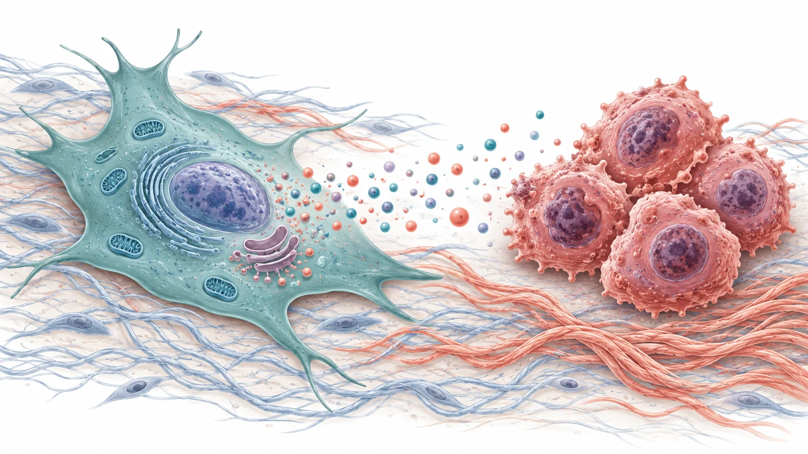
The review describes two main routes by which senescent MSCs may influence a tumor: through released molecules and through physical remodeling of the extracellular matrix, the network of material surrounding cells.
The secreted route is illustrated by experiments using conditioned medium, the liquid collected after MSCs have been grown in culture. In one breast-cancer example, medium from senescent umbilical-cord MSCs increased proliferation and migration of MCF-7 and MDA-MB-231 cancer cells in laboratory tests. The review identifies increased interleukin-6, or IL-6, and activation of STAT3 in the cancer cells as a proposed explanation for those observations. In a xenograft experiment described by the review, combining breast-cancer cells with senescent MSCs was associated with larger and more vascularized tumor nodules.
The important point is not that every senescent MSC releases a universal tumor-promoting mixture. SASP composition varies with the cell source, the stress that induced senescence and the surrounding conditions. IL-6 is highlighted because multiple studies associated it with cancer-cell proliferation, movement and resistance to chemotherapy, but it operates within a much broader and changing secretory environment.
The physical route is visible in a three-dimensional model containing MSCs, breast-cancer cells and a surrounding matrix. Senescent MSCs changed their production of matrix components, inhibitors of matrix-degrading enzymes and crosslinking agents. The resulting structure influenced how breast-cancer cells moved out of spheroids and how readily they proliferated. The review also suggests that matrix remodeling and soluble factors from senescent MSCs may increase the motility of nearby pre-senescent MSCs.
A cell that has stopped dividing can therefore remain consequential without becoming a cancer cell itself. Its continuing output can change the chemical conversation around a tumor, while its altered construction of the surrounding matrix can change the physical routes available to cancer cells.
The same secretome can meet different cancers
The review also makes clear why a simple rule about senescent MSCs would be misleading. In another set of experiments, secretions from senescent adipose-tissue MSCs promoted senescence or apoptosis in ARH-77 multiple-myeloma cells and reduced the number of cells positive for Ki-67, a marker used in the reported experiments to identify actively cycling cells.
The response changed after the MSCs had been exposed to myeloma cells. Following that interaction, the senescent adipose-tissue MSCs no longer showed the same ability to reduce the cycling myeloma-cell population. The cancer cells had apparently altered the stromal secretory environment enough to remove the observed inhibitory effect.
The prostate-cell comparison points to another source of variation. In experiments using acute senescence induced by hydrogen peroxide or low-dose X-ray exposure, senescent MSC secretions stimulated senescence and reduced proliferation in immortalized PTN2 prostate cells. The same treatment did not affect PC3 metastatic prostate-cancer cells in the reported model.
These examples describe tested cell systems, not a general ranking of cancers by sensitivity. They show that the outcome can depend on the cancer cell’s state, its prior interaction with stromal cells and the way senescence was induced. A secretion that inhibits one tested model may have little effect on another, while a tumor-promoting response can emerge when the surrounding cells have been re-educated by cancer signals.
Why the experiments are hard to interpret
MSCs are difficult to study in their natural setting. They are relatively scarce in tissues, so researchers often expand them in culture. Yet prolonged expansion can itself produce replicative senescence. An experiment may therefore compare cells that are genuinely altered by an aging organism with cells that have acquired some senescent features because of the laboratory process used to obtain enough of them.
The review notes that gene-expression patterns in MSCs from older donors can resemble those of MSCs made senescent in vitro, which makes cultured cells useful for investigation. Even so, similarity does not erase the experimental distinction. A culture model can reveal how a secretome affects a cancer-cell line under defined conditions, but it cannot by itself establish how common that interaction is inside a human tumor or how large its effect would be.
The direction of influence can also run both ways. Prostate-cancer secretomes changed co-cultured MSCs, increasing the release of inflammatory factors while reducing MSC proliferation and migration. MSCs taken from people with multiple myeloma showed a senescent profile with altered size, growth, differentiation potential, immune-regulating capacity and secreted proteins. These observations indicate that tumors can reshape surrounding stromal cells, but they do not by themselves show which changes drive tumor behavior or what portion of the relationship matters clinically.
Cancer treatment adds another possible source of stress. Chemotherapy and radiation can cause persistent DNA damage and senescence in surrounding MSCs. The review presents this as one possible contributor to a microenvironment that allows tumor cells to persist or that may support later malignancy. It remains a mechanistic concern from the reviewed evidence, not a patient-level estimate of cancer risk.
Several therapeutic levers, none yet settled
The therapeutic discussion separates several strategies that are often grouped together but would do different things.
Senolytics are intended to eliminate senescent cells by disabling survival pathways that protect them from apoptosis. In MSC-related experiments, quercetin improved some measures of older rat MSC function and, with dasatinib, improved osteogenic capacity in aged mouse MSCs. Navitoclax reduced the proportion of senescent mouse or human bone-marrow MSCs in the reported studies, but it also reduced osteogenic potential in one mouse model and did not restore some markers of cellular youth in human cells.
Those findings come mainly from regenerative or age-related models. Improvements in bone formation, angiogenesis or cardiac repair do not demonstrate that a strategy treats cancer. They do, however, illustrate the central safety problem: removing or altering senescent MSCs may also remove functions that tissues need, or may change repair capacity in an undesirable direction.
A second approach is to modify the SASP rather than eliminate every senescent cell. Senomorphic strategies would aim to reduce harmful chronic inflammatory signals while preserving useful immune or repair activity. The review describes work targeting amphiregulin, an EGFR ligand associated with an immunosuppressive tumor microenvironment. In a mouse prostate-cancer model treated with genotoxic therapy, targeting this factor was associated with reduced chemoresistance and restored immune competence. This is bounded animal evidence about one signaling component, not proof of a general MSC-targeted cancer treatment.
A third direction is to restore selected functions in aged or senescent MSCs. Experimental overexpression of molecules such as NDNF, apelin or MIF improved particular MSC properties in laboratory or cardiac-repair models. These results may help researchers understand how senescence is regulated, but the reported regenerative outcomes are not evidence of anticancer efficacy.
The review also presents a proposed “two-punch” strategy. Cancer cells could first be pushed into senescence to limit their division, followed by senolytic treatment to remove senescent cancer and stromal cells. In principle, that sequence could combine a growth-arresting effect with clearance of cells whose later secretions might support the tumor. In practice, the proposal raises a difficult targeting problem: treatment would need to distinguish the intended cancer cells and harmful stromal cells from cells whose senescence or repair functions should be retained. The reviewed evidence does not establish this combined regimen as safe or effective.
What this could change for cancer treatment
The most useful future capability would be an adjunct treatment that reduces tumor-supporting signals and drug resistance arising from the surrounding tissue, rather than targeting cancer cells alone. The review’s 2021 perspective suggests that this might involve selectively removing harmful senescent cells, adjusting their secretions or changing specific stromal functions while preserving beneficial immune and repair roles.
Patients could benefit if such targeting proves effective and safe in the relevant tumors. Reaching that point would require showing that the intervention works in the right cellular context, avoids damaging useful tissue functions and produces a meaningful cancer benefit beyond the experimental models described here. The promise lies in making the tumor’s surroundings less supportive; the unresolved challenge is doing so selectively.
Key Sources
- Primary source: Tumorigenic Aspects of MSC Senescence-Implication in Cancer Development and Therapy.
DOI or Journal Page · Review or Meta-Analysis · 2021-11-02
Keep exploring