
Research Signal – Source date 2021-10-18
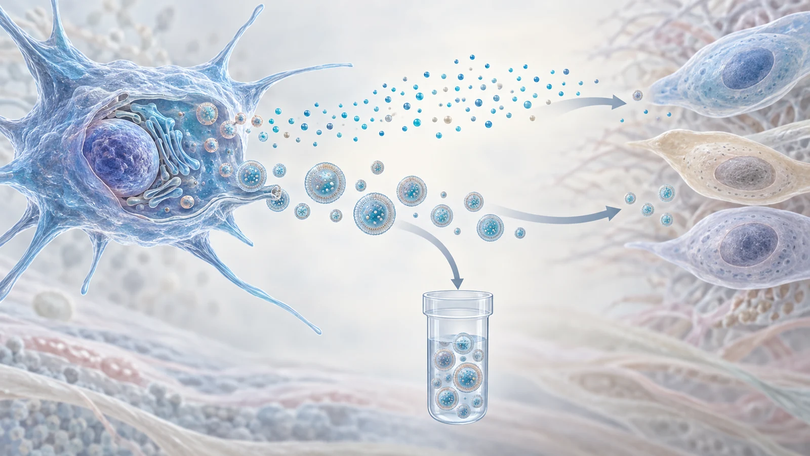
Mesenchymal stem cells, or MSCs, are adult cells that can form bone, cartilage and fat. That makes them appealing in conditions where those tissues lose function. But the most important question may not be whether transplanted cells permanently replace damaged ones. It may be whether they briefly change the environment around an injury or an aging tissue by releasing useful biological messages.
A review published on 18 October 2021 examined that possibility across ordinary physiological aging, premature-aging syndromes, and specific age-related conditions. Its clinical examples included knee osteoarthritis and frailty. Its broader argument was that MSCs may act less like replacement parts and more like temporary signal sources. The review also considered whether those signals could eventually be collected in cell-free products made from extracellular vesicles, tiny membrane-bound packages released by cells.
Aging can weaken the cells meant to support repair
Aging affects the regenerative capacity of tissues through several interacting changes, including altered communication between cells, cellular senescence and exhaustion of tissue-specific stem-cell reserves. MSCs are part of that story. As they become senescent, they proliferate and migrate less effectively, their ability to differentiate into specialized cells is impaired, and their secretory behavior changes.
Those secretions matter because senescent MSCs can acquire a senescence-associated secretory phenotype, or SASP. The review describes this as a mixture that includes pro-inflammatory cytokines, matrix-remodeling enzymes, reactive oxygen species and chemotactic molecules. Released locally, these factors can influence neighboring cells and encourage further senescence through local cell-to-cell effects. A cell that once helped maintain tissue may therefore become less capable of supporting repair while contributing to more inflammatory surroundings.
The result is not simply fewer new cells. It is a change in the tissue environment in which cells must survive, communicate and respond to injury. That helps explain why the review focuses on both the condition of the administered MSCs and the messages they release.
The proposed therapy may work through communication
MSCs can migrate toward damaged tissue and, in some circumstances, differentiate into specialized cell types. Yet the review presents their main therapeutic effect as potentially paracrine. In plain language, paracrine signaling means that one cell releases bioactive factors that influence nearby cells without needing to become those cells itself.
This interpretation is partly based on observations that beneficial effects can occur even when MSC engraftment in a target tissue is limited. The review describes the action as a possible “hit-and-run” mechanism: administered cells may encounter an injured or inflamed environment, release a changing mixture of factors, and then disappear or remain only briefly while the surrounding tissue has already received a biological instruction.
That proposed mechanism shifts the development question. Researchers are not only asking whether enough cells reach a target and survive there. They are also asking what those cells secrete, how the recipient environment shapes that secretion, and whether the useful components can be separated from the living cells themselves. In the 2021 review, this was a mechanistic interpretation supported by a mixture of cell studies, animal work and clinical observations, not a universal explanation established for every MSC preparation.
What the human studies actually measured
The clinical examples in the review concern particular problems rather than a general treatment for aging. Knee osteoarthritis is one example. Phase I trials summarized in the review reported that intra-articular administration of autologous MSCs, taken from the same person, or allogeneic MSCs, obtained from a donor, was safe in those studies and associated with reductions in pain and inflammation alongside functional improvements.
The comparisons also raised a question about the age of the starting cells. In the summarized single-administration studies, autologous MSC treatment was not as efficient as allogeneic treatment, and higher cell amounts were used in some autologous comparisons. A lower amount of allogeneic MSCs was reported to produce similar clinical improvements in the cited trials. These cross-study dose comparisons do not establish that allogeneic cells are universally better or that one dose is optimal; the preparations, protocols and participants were not interchangeable.
A randomized phase I/II trial examined two intra-articular doses of allogeneic umbilical-cord MSCs, six months apart, alongside hyaluronic-acid treatment. The review reports that this repeated-dose approach was safe and clinically superior to a single MSC dose in that study. The transient benefit seen after some single-dose treatments, together with low retention and survival at the injection site, was consistent with a paracrine contribution. It did not show that living MSCs rebuilt an entire aging joint or reversed the biology of aging.
Frailty provides a different clinical context. It is a syndrome in which age-related deterioration across physiological systems leaves a person less resilient to stress. The review describes two successive CRATUS trials of allogeneic bone-marrow MSCs from relatively young donors. In the first, a small, nonrandomized dose-escalation study, one intravenous infusion of 20, 100 or 200 million cells was given to five people in each group; the participants had a median age of 78.4 years. Outcomes were assessed one, three, six and twelve months after infusion.
The treatment was reported as safe and well tolerated in that study. All dose groups increased their six-minute walk distance at three and six months, while the 100-million-cell group showed the greatest improvement in the physical component of quality-of-life questionnaires, beginning at the first month. These were functional and patient-reported measures in an older group receiving a particular cell preparation. They were not measurements of restored biological youth, lifespan extension or a broadly established healthspan effect.
Premature aging shows how stem-cell dysfunction develops
Rare progeroid disorders offer researchers a concentrated way to study processes that also have relevance to aging biology. In the models reviewed, abnormal forms of nuclear-lamina proteins, including prelamin A and progerin, were introduced into human MSCs or examined in MSCs derived from patients’ induced pluripotent stem cells.
Those models connected the abnormal proteins with senescence and impaired MSC function. The cells showed problems with adipogenic and osteogenic differentiation, as well as changes involving autophagy, SASP and viability under stress. In adipogenic models, abnormal prelamin A accumulation was associated with mitochondrial dysfunction, endoplasmic-reticulum stress and altered lipid metabolism. The review also describes reduced therapeutic potential when affected cells were transplanted into a mouse ischemic hind-limb model.
These experiments help explain how a defect in nuclear architecture can disrupt the behavior of a mesenchymal stem-cell population. They are models of premature aging and cellular dysfunction, however, not proof that a therapy tested in those systems would treat ordinary aging in people.
The review mentions a case report involving a child with Hutchinson–Gilford progeria syndrome who received two infusions of stromal vascular fraction isolated from the patient’s mother. The material was a heterogeneous mixture that included MSCs, rather than a defined MSC product. That difference matters: an observation after a mixed-cell preparation cannot establish an effect attributable specifically to MSCs.
Vesicles turn a living-cell idea into a product question
Extracellular vesicles are membrane-bound packages released by cells. They can carry biological material from the cell that produced them, allowing researchers to investigate whether some of the effects associated with MSCs could be delivered without administering living cells.
The review describes strong source- and age-dependent differences in vesicle behavior. Vesicles from induced pluripotent stem cells were reported to be produced in greater amounts than those from neonatal or adult MSCs, while neonatal MSCs produced more than adult MSCs in the cited work. Vesicles from young MSCs also showed stronger immunomodulatory activity than vesicles from older MSCs, a difference associated with variation in their molecular cargo.
The evidence is not limited to MSC-derived vesicles, and the sources should not be blended together. Experiments with vesicles from young fibroblasts reduced markers of senescence and improved proliferation in cultured elderly human fibroblasts and fibroblasts from a progeria model. In aged mice, injections of those young fibroblast-derived vesicles reduced senescence markers in several tissues. These findings concern cell behavior and animal markers, not demonstrated clinical benefit from MSC-derived vesicles in older people.
The review also discusses findings involving vesicles from young umbilical-cord MSCs in aged mouse models. Together, these studies made cell-free treatment an attractive direction, but they left the product itself incompletely defined. Researchers still needed to establish reliable ways to characterize vesicles, identify which molecules drive the observed effects, and determine which cell populations receive and respond to their cargo. A package that varies with its source cell and donor age cannot be treated as a standardized medicine merely because it is called an extracellular vesicle.
A cell treatment is shaped by its starting material and its setting
The identity of the cell is only one part of an MSC treatment. The review emphasizes the age of the donor and the number of times cells have been expanded in culture. Both affect the youthfulness and functional properties of the starting material. A preparation made from young cells may therefore differ biologically from one made from cells that have aged in the body or undergone prolonged culture.
The cells can also respond to conditions before administration. Researchers have explored “priming,” “licensing” or “preconditioning,” in which culture conditions are adjusted to resemble aspects of the environment the cells will encounter. Examples discussed in the review include low-oxygen conditions and exposure to molecules associated with injured or inflamed tissue. Interferon gamma, for instance, was used to enhance immunosuppressive properties even in senescent MSCs, alongside changes in proteins involved in that response.
The recipient environment introduces another variable. Immune status, hypoxia, inflammation, extracellular-matrix changes and disease severity can all influence what administered MSCs do. Young cells placed into an older biological setting may also be affected by age-related factors in the surrounding serum. The practical implication is not that the review supplies a ready-made recipe for treatment. It is that cell source, culture history, preparation conditions and recipient biology interact, making a living-cell therapy difficult to define by cell name alone.
Why cell-free therapies could matter for older adults
For older adults whose mobility or resilience is impaired, the most useful future outcome would not be a vague promise of rejuvenation. It would be a reliable way to support the tissues and physiological functions that aging has made harder to maintain. If researchers can identify the beneficial messages released by suitably prepared MSCs and package them in a consistent cell-free product, treatment might eventually be easier to characterize, store and administer than a living-cell product.
That possibility remains conditional from the review’s 2021 perspective. The active cargo, target cells, manufacturing characteristics, clinical safety and patient benefit would all need to be demonstrated. A defined vesicle-based treatment is therefore a development prospect, not an established therapy for healthy aging. Its significance lies in the specific capability it could offer: delivering a controlled biological message without requiring transplanted cells to survive and function in exactly the right place.
Key Sources
- Primary source: Cell and Cell-Free Therapies to Counteract Human Premature and Physiological Aging: MSCs Come to Light.
DOI or Journal Page · Review or Meta-Analysis · 2021-10-18
Keep exploring