
Research Signal – Source date 2021-05-18
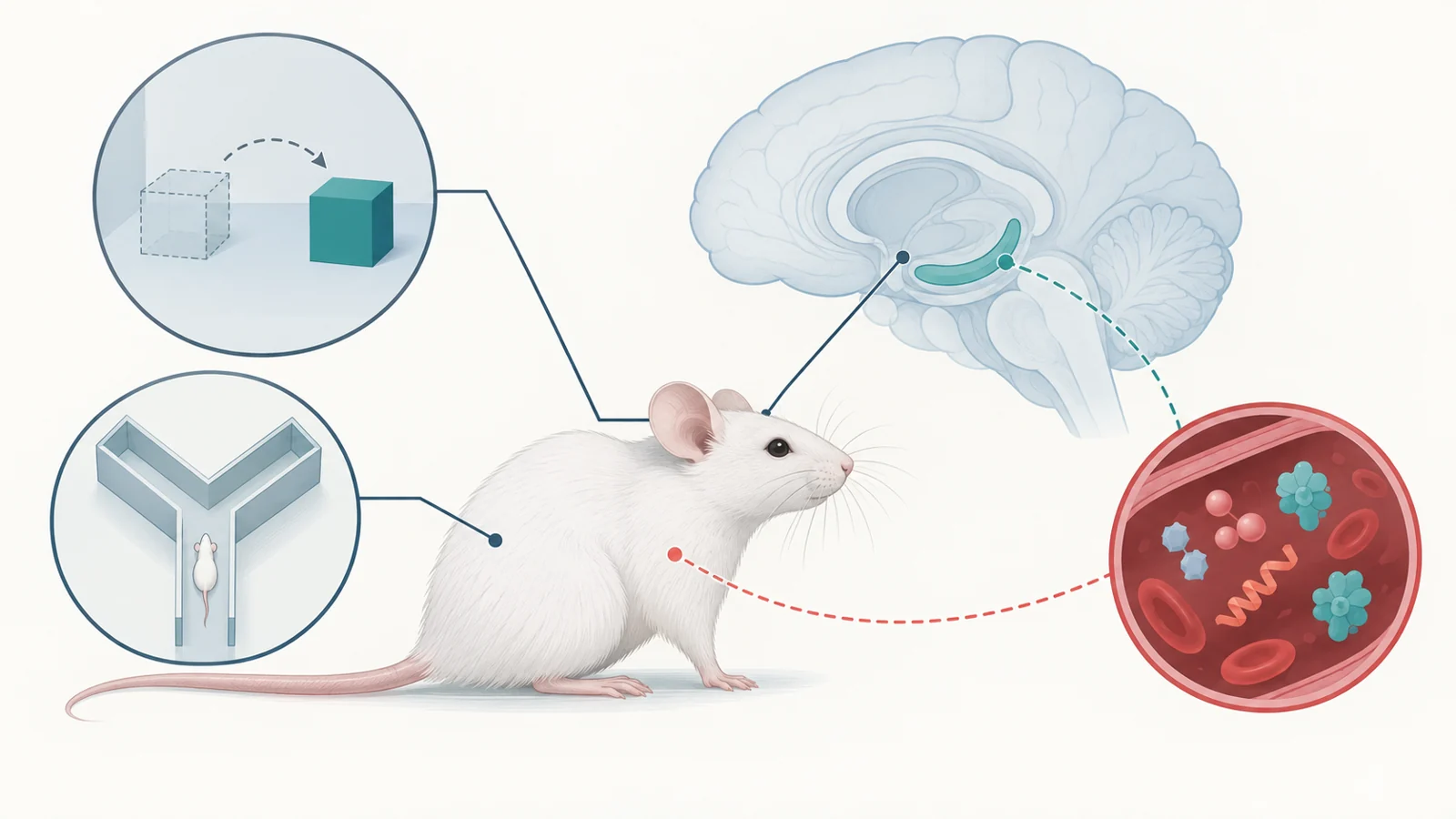
Prolonged stress can affect behavior and memory, but an association alone does not show which biological changes matter. A study published on May 18, 2021 asked whether cellular senescence—a stable stress- or damage-associated state in which cells stop dividing—might be a modifiable contributor. In young male mice exposed to chronic unpredictable stress, dasatinib plus quercetin improved performance on two spatial-memory tasks after the stress period. The treatment also reduced senescence-associated measurements in the hippocampus and some blood-borne signaling proteins.
The result gives researchers a reason to investigate senescence as a contributor to stress-related memory problems. It does not show that brain aging was reversed, or that the same treatment would help people. Its value lies in the experiment’s sequence: stress came first, and a drug intervention followed.
A month of unpredictable stress changed two memory tasks
The researchers used eight-week-old male C57BL/6 mice. Over one month, the animals were exposed to chronic unpredictable stress, or CUS, through a rotating set of conditions that included food or water deprivation, altered light and dark periods, crowding, cage tilting, restraint, temperature changes, social defeat, sleep deprivation and other disruptions. The mice were randomly subjected to one form of stress each day.
After this period, the CUS-treated mice showed depression-like behaviors and performed worse than nonstressed controls on spatial-memory tests. The study did not treat cognitive decline as one general score. It used two specific tasks that ask mice to notice a change in familiar space.
In the object-location test, mice first explored two identical objects. One object was moved to another position 24 hours later. The researchers measured how much attention the animals gave to the object in its new location compared with the familiar one. In the Y-maze test, one arm was closed during an initial exploration period and opened one hour later. Preference for the newly available arm provided another measure of spatial memory.
The authors report that roughly 80% to 90% of CUS-exposed mice developed the behavioral pattern used for the study’s senescence analyses. Some stressed mice did not show the same depression-like behavior and memory decline. The researchers confirmed the behavioral changes before examining senescence-associated measurements, which helped connect the biological analysis to the animals that had displayed the relevant response.
The hippocampus showed several signs of senescence
Cellular senescence is a stable state associated with injury or stress. Senescent cells stop dividing and can release signaling molecules known as senescence-associated secretory phenotype, or SASP, factors. Senescence is not automatically harmful: the state can participate in repair. The question in this study was whether stress-related accumulation or activity of senescent cells might contribute to memory impairment.
The researchers used more than one measurement because each captures a different part of the question. Senescence-associated beta-galactosidase, or SA-beta-gal, staining identifies enzyme activity associated with senescent cells in tissue. In the stressed mice, SA-beta-gal-positive cells were especially apparent in the dentate gyrus, a hippocampal region involved in the tested memory behaviors. The researchers also measured hippocampal p16INK4a protein, another senescence-associated marker, and found higher levels after CUS exposure.
These measurements were related to performance. Higher SA-beta-gal activity and higher hippocampal p16INK4a were associated with poorer results in the object-location and Y-maze tasks. The pattern is consistent with senescence accompanying the memory deficit, but a stain or protein measurement is not a direct reading of brain age and does not by itself explain how memory changed.
The study also examined which tested brain cell markers overlapped with SA-beta-gal-positive cells. The staining colocalized with NeuN, a neuronal marker, but not with the tested markers for astrocytes, microglia or oligodendrocytes. That observation narrows the cellular location of the measured signal in this experiment. It still does not establish that selective clearance of a particular hippocampal cell population caused the behavioral outcome.
A post-stress treatment tested whether the signal was modifiable
The intervention came after the mice had undergone CUS. The researchers gave dasatinib plus quercetin, a drug combination used in the experiment as a senolytic treatment, by oral gavage five days a week for two weeks. The stressed mice were compared with stressed mice given vehicle rather than the drug combination.
After treatment, the D plus Q group had fewer SA-beta-gal-positive cells and lower hippocampal p16INK4a measurements. Drug-treated CUS mice also performed better on the object-location and Y-maze tasks than vehicle-treated CUS mice. The improvement was therefore not just a correlation observed at one time point: a post-stress intervention changed both the senescence-associated measurements and the memory-task results.
The experiment included an important control for a simpler explanation. In an open-field test, travel distance and movement velocity did not differ significantly across the reported treatment comparisons. That makes it less likely that the memory-test difference was simply caused by a broad change in locomotion or the ability to explore. It does not identify which cells or molecular route produced the benefit, and it does not show that the drugs acted only in the hippocampus.
Blood measurements widened the question
The researchers also measured signaling proteins in serum. CUS increased several SASP-related factors, including interleukin-1 beta, interleukin-6 and interleukin-13. D plus Q treatment reduced these measurements. Higher levels of each were associated with poorer performance on the spatial-memory tests.
Those findings raise the possibility that stress-related senescence could influence memory through signals outside the hippocampus as well as through local brain changes. A circulating factor could reflect activity in multiple tissues, and a blood measurement can change alongside behavior without being the cause of that behavior. The study’s correlations do not establish that these proteins carried the treatment benefit, nor do they identify their cellular source.
This matters because the intervention was systemic. Reduced serum signaling and improved memory occurred together with reduced hippocampal senescence-associated measurements, but the design does not separate those effects into a single causal chain. The treatment comparison supports further investigation of senescence and its secreted factors; it does not settle which part of that biology is necessary for the behavioral improvement.
What the mouse study can and cannot establish
The study’s population was narrow: young male mice of one strain exposed to a particular rotating pattern of stressors. The response may differ with age, sex, genetic background or another form of prolonged stress. The fact that some CUS-exposed mice did not develop the measured behavioral pattern also points to variation within the model rather than a uniform response to stress.
The safety observations were limited as well. The researchers measured selected serum indices related to liver and kidney function. Some values did not differ across the reported groups, while another liver-related measure changed with stress and treatment. These checks provide context for the experiment, but they are not a comprehensive or long-term safety assessment. Dasatinib is also an anticancer drug, so a result in mice cannot be turned into self-treatment guidance for memory problems.
Two improved spatial-memory tasks are meaningful within this model, but they do not establish reversal of brain aging, prevention of dementia or restoration of every aspect of cognition. The evidence presented here also does not establish how long the improvement persisted after treatment. Moving from this mouse experiment to a human intervention would require evidence that the relevant biology operates in people and that targeting it produces a meaningful benefit with acceptable safety.
What this could mean for stress-related memory problems
The study’s most useful implication is a conditional one. If prolonged stress produces senescence-related changes that help sustain memory problems, treatments directed at senescent cells or the signals they produce could eventually offer a way to help people retain or regain memory function affected by prolonged stress. That possibility depends on reproducing the biological relationship in people, identifying an intervention that acts through a meaningful route and establishing acceptable safety over the relevant period.
The 2021 experiment does not provide that human outcome. It provides a treatment comparison that gives researchers a reason to keep testing whether senescence is a modifiable contributor to stress-related memory difficulties.
Key Sources
- Primary source: Cellular senescence as a driver of cognitive decline triggered by chronic unpredictable stress.
DOI or Journal Page · Peer-Reviewed Paper · 2021-05-18
Keep exploring