
Research Signal – Source date 2021-01-01
Aging creates an apparent contradiction in the brain. Experience can continue to reshape connections between neurons, yet the systems that support those connections may become less reliable over time. Memory therefore depends on more than the brain’s ability to change. It also depends on energy production, waste processing, communication between neurons and support cells, and the way stressed cells influence their neighbors.
A review published in January 2021, “Mechanisms of neuroplasticity and brain degeneration: strategies for protection during the aging process,” brings these threads together. The authors searched PubMed and preferentially used references from 2015 to 2019, while retaining older sources when classical information was needed. The review connects the biology of neuroplasticity with processes that may contribute to neuronal damage, then discusses exercise, environmental enrichment and other protective strategies. Its evidence spans cellular mechanisms, animal experiments and human observations, so the findings answer different questions rather than forming one test of a single memory-preservation intervention.
Why an adaptable brain can still become vulnerable
Neuroplasticity is the brain’s continuing ability to alter its structure and function in response to activity. At the most tangible level, this means that synapses, the specialized junctions through which neurons communicate, can be strengthened, weakened, renewed or reorganized. Learning and memory are associated with these activity-dependent changes. The brain’s adaptability is therefore not an abstract property floating above biology. It is built from repeated adjustments in molecular signaling, synaptic structure and neuronal circuits.
One process discussed in the review is long-term potentiation, an electrophysiological change associated with a lasting increase in synaptic efficacy. It can accompany changes in gene transcription and protein synthesis, helping stabilize structural and functional alterations in a connection. Plasticity includes both structural changes, such as dendritic spine formation and the generation of new neurons, and functional changes, such as altered neurotransmitter release, receptor sensitivity and postsynaptic signaling. New-neuron formation is part of this picture, but it is not the whole meaning of an adaptable brain. Much of the ongoing work occurs through the adjustment of connections that already exist.
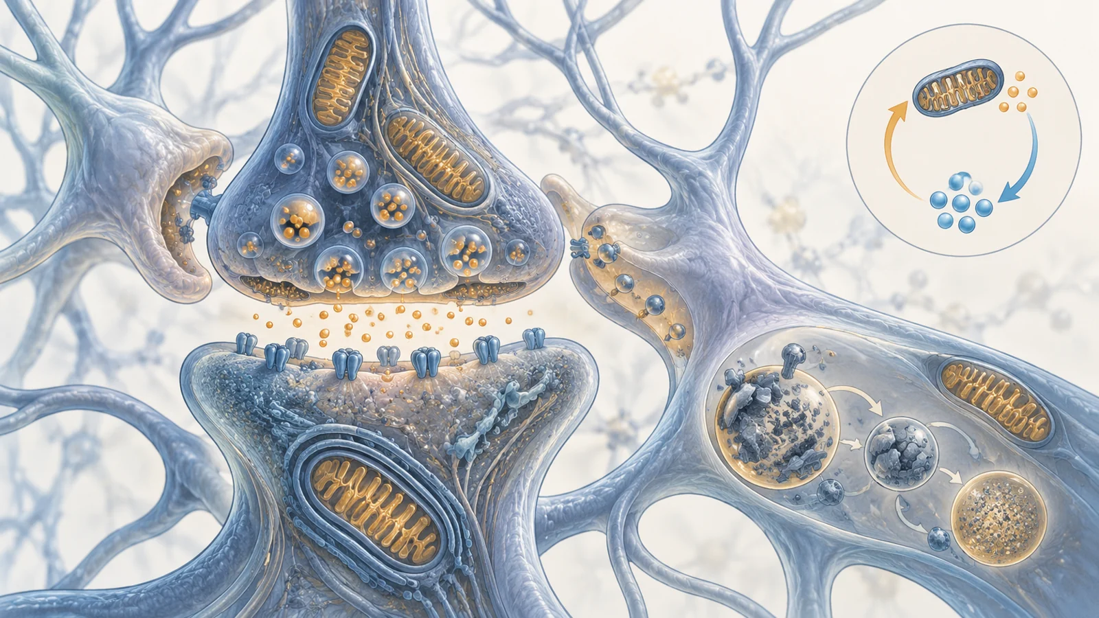
Astrocytes also participate in this process. They are positioned around synapses in what is often called a tripartite synapse, alongside the presynaptic and postsynaptic parts of the neuronal connection. Their processes can help stabilize and mature dendritic spines, detect synaptic activity and influence how strongly a connection operates. Astrocytes also take up substances including glutamate from the space around the synapse, helping limit excessive stimulation that could become harmful.
Brain-derived neurotrophic factor, or BDNF, is another part of the review’s account. The protein is involved in synaptic changes and supports processes related to both structural and functional plasticity. The review also connects BDNF with neurogenesis in the dentate gyrus. These mechanisms make adaptability possible, but they require continuous cellular maintenance. A connection cannot keep responding to experience if the neuron lacks energy, accumulates damaged components or remains surrounded by persistent inflammatory signals.
Energy and calcium can push neurons toward trouble
Mitochondria are central to this maintenance problem because they supply much of the cell’s usable energy. The review notes that approximately 90% of ATP production occurs in mitochondria and that mitochondrial dysfunction is associated with neurodegenerative disease, reduced ATP production, problems in the electron transport chain, increased reactive oxygen species and changes in mitochondrial DNA.
Mitochondria also help regulate calcium. Calcium ions participate in metabolic signaling and neuronal function, and mitochondria can buffer them after synaptic activity. This buffering helps preserve neuronal polarity and supports structural adaptations in dendritic spines. Calcium is therefore not simply a damaging substance to be removed. It is part of normal signaling whose concentration and timing must remain controlled.
The review describes a feedback sequence that helps explain why neuronal resilience can weaken. When mitochondria produce too little ATP, energy-dependent calcium pumps in the plasma membrane and endoplasmic reticulum may work less effectively. Calcium can then accumulate inside the cell. Excess calcium can activate pathways associated with cell death, while oxidative stress can further reduce the mitochondria’s ability to generate energy. Calcium entering mitochondria may also overload their proton circuit and contribute to a loss of mitochondrial permeability control.
This sequence is important because it shows why simply increasing brain activity is not a complete account of protection. Activity can help shape synapses, but signaling also depends on the cell’s capacity to supply energy, buffer calcium and recover from metabolic stress. Plasticity is an active balancing process, not an unlimited reserve that operates independently of the machinery keeping neurons alive.
Recycling keeps damaged cell parts from accumulating
Autophagy is the cell’s breakdown and recycling process. Through lysosomal machinery, it helps remove and digest damaged proteins, organelles and other cellular contents, then recycles usable components. The process is needed under ordinary conditions as well as during stress. Neurons are especially dependent on reliable internal cleanup because dysfunctional proteins cannot simply be diluted through frequent cell division.
The review uses Beclin1 to show how different kinds of evidence can illuminate this system. A reduction in Beclin1 was observed in the brains of people with Alzheimer’s disease. That observation is relevant to the disease state, but it does not by itself show whether impaired autophagy caused the neuronal changes or how altering the pathway would affect memory in people.
The experimental evidence described alongside it comes from PDAPP mice. Removing the Beclin1 gene compromised autophagy and was associated with greater accumulation of intraneuronal beta-amyloid, reduced neuronal autophagy, neurodegeneration, lysosomal rupture and microglial alterations. Increasing Beclin1 in that experimental context reduced intracellular and extracellular amyloid. These mouse manipulations help test the consequences of changing one part of the pathway, while the human observation and the animal experiment remain distinct forms of evidence.
Autophagy is also a sequence of coordinated steps rather than an on-off switch. The review emphasizes that induction, maturation and clearance all need to be regulated. A problem at any stage could leave cellular material incompletely processed. For that reason, the findings support interest in autophagy as a neuroprotection target, but they do not establish that broadly increasing recycling would safely improve human memory or prevent dementia.
Activity findings depend on what was measured
The review discusses physical activity and environmental enrichment as ways of engaging protective mechanisms, including pathways involving BDNF and other signals. The examples are informative partly because they differ in species, duration and outcome.
In one animal example, moderate physical activity over 11 weeks improved the cognitive performance of rats that had been less responsive to a memory task. A separate study cited in the review associated moderate activity with neurogenesis, protection from neuronal death and neuronal differentiation, whereas intense activity did not produce similar effects. These findings come from different experiments and do not establish a universal prescription for intensity or duration in people.
Environmental enrichment supplied a different kind of animal evidence. In a transgenic mouse model of Alzheimer’s disease, an enriched environment was associated with better memory retention. Other work discussed in the review linked environmental improvement with lower levels of the pro-inflammatory cytokine IL-1 beta and an increase in astrocytes. These findings suggest that surroundings and activity can influence several biological layers at once, but the mouse model and its measured outcomes cannot be treated as a direct forecast of human daily function.
The human examples in the review raise a separate timing issue. Interventions lasting 12 or 16 weeks did not significantly change some measured parameters related to cognition, including cerebral blood flow or growth factors. In contrast, people with higher activity over a longer period of one year were reported to have greater hippocampal volume. A growth-factor measurement, a blood-flow measurement, a brain-volume measurement and a cognitive test do not answer the same question. The differences make it impossible to extract a single minimum exercise duration from these examples, or to assume that a change in hippocampal volume automatically represents lasting improvement in learning and memory.
The practical implication at the review’s 2021 vantage point is not that one activity schedule had been proven to preserve cognition. It is that duration, biological response and functional outcome need to be considered together. The evidence supports continued investigation of active and stimulating environments while leaving the size, timing and durability of any human benefit open.
Senescent cells create a difficult target
Cellular senescence is a state of essentially irreversible cell-cycle arrest that can arise through mechanisms including telomere shortening, oncogene activation, oxidative stress and cell-to-cell fusion. Senescent cells can release a collection of signaling molecules known as the senescence-associated secretory phenotype, or SASP. These secretions include inflammatory cytokines, chemokines, growth factors and proteases that can alter neighboring tissue.
In the brain, the concern is not limited to the senescent cell itself. The inflammatory environment created by its secretions may affect nearby neurons and glial cells. The review discusses links between tau phosphorylation and SASP release, as well as findings in which amyloid-beta plaques were associated with senescent behavior in oligodendrocyte progenitor cells. These examples point toward a possible route by which cellular aging and protein-related pathology could reinforce a damaging local environment.
Senolytics are agents intended to eliminate senescent cells. They are appealing because removing cells that release harmful signals might reduce some of the surrounding stress. Yet the review stresses a central difficulty: not every senescent cell is necessarily harmful in every context. Senescent cells can participate in useful processes such as wound healing. A strategy that removes them indiscriminately could therefore interfere with functions that the body still needs.
The animal-to-human boundary is especially important here. The review describes growing interest in potential senolytic substances, but the in vivo evidence it discusses comes from animal studies with limited translational correlation to human biology. The long-term effects also require investigation. At the review’s publication date, senolytic research was a direction for testing how cellular aging might be modified, not evidence that eliminating senescent cells safely protects human memory.
The same caution applies to compounds sometimes described as natural senolytics or neuroprotective agents. Their appearance in a research discussion does not make them proven therapies, and the supplied evidence does not support treating plant-derived compounds as established ways to preserve cognition. The unresolved question is not merely whether senescent cells can be reduced. It is whether harmful signaling can be limited while useful cellular roles remain intact, and whether that balance produces a durable functional benefit in humans.
What this could mean for memory in later life
The review presents aging-related neurodegeneration as a problem of interacting systems. Synapses must remain adjustable, astrocytes must help regulate their surroundings, mitochondria must supply energy and manage calcium, and autophagy must process damaged material. Senescent cells add another layer because their secretions can influence the tissue around them while their presence may still serve beneficial purposes in some settings.
For older adults, the prospective benefit is concrete but conditional. Approaches that sustain synaptic function and reduce damaging cellular processes might help preserve the ability to learn, remember and manage familiar tasks, potentially supporting everyday independence. Reaching that outcome would require durable human functional benefits, not just a molecular change, a larger measured brain region or an encouraging result in an animal model. It would also require interventions that protect useful cellular functions rather than treating every stress response as something to eliminate.
The January 2021 review therefore offers a way to understand why memory protection is scientifically difficult. The brain remains capable of change, but that capacity depends on upkeep across several linked systems. Exercise, stimulating environments, metabolic support, autophagy and senescent-cell targeting may each illuminate part of the problem. None, in the evidence summarized here, establishes a reliable method for preventing or reversing dementia in humans.
Key Sources
- Primary source: Mechanisms of neuroplasticity and brain degeneration: strategies for protection during the aging process.
DOI or Journal Page · Review or Meta-Analysis · 2021-01-01
Keep exploring