
Research Signal – Source date 2026-06-01
Surviving sepsis, serious trauma, burns, or major surgery can mark the beginning of another difficult phase: trying to think clearly again. Some people recover from the immediate medical emergency but continue to struggle with attention, memory, planning, or the mental energy needed for ordinary tasks.
A June 1, 2026 review, Neurocognitive Aging Following Acute Illness: Pathobiology and a Framework for Developing Neurotherapeutic Agents, brings together clinical observations and mechanistic research on that problem. It is a narrative, focused review rather than a treatment trial. Its clinical-study coverage of symptoms after critical illness extends through December 2025, while its larger purpose is to organize possible biological targets for future neurotherapeutic drugs.
The review cites estimates that 50%–70% of patients experience impairments associated with post-intensive-care syndrome, which encompasses cognitive, psychological, and physical difficulties, with problems reported years after discharge. Those figures describe a broad and varied group, not a single predictable outcome. The central question is how an illness that begins elsewhere in the body might leave the brain less able to support memory, attention, and everyday independence.
The first signal starts outside the brain
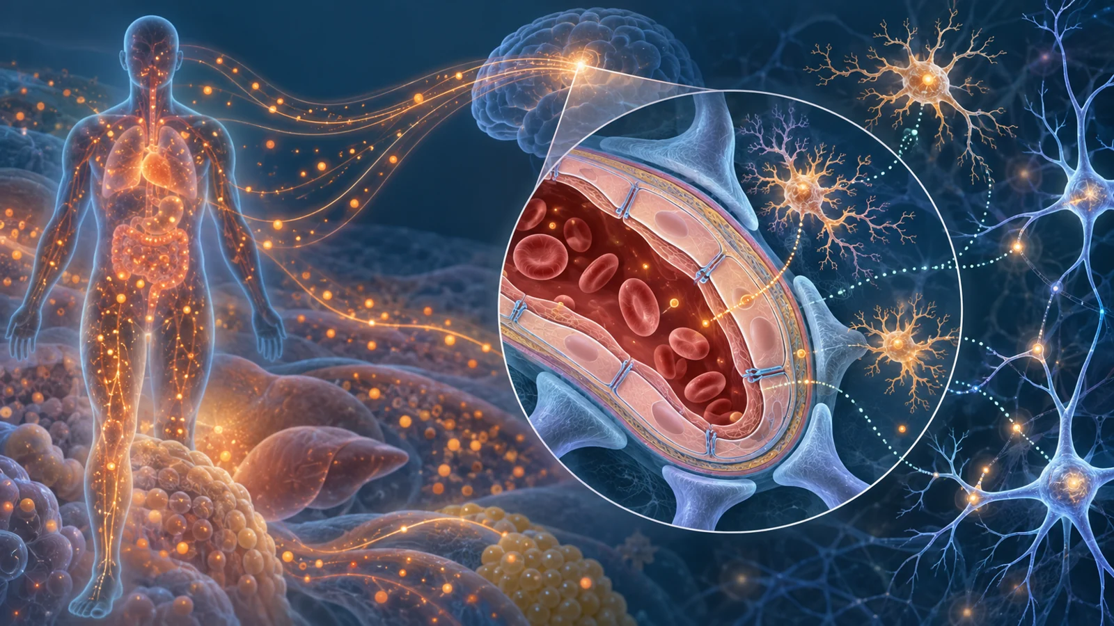
Acute systemic inflammation is the body’s rapid immune mobilization after infection, trauma, or major surgery. It is meant to contain danger and support repair. Immune cells, blood-vessel lining cells, and other tissues release signaling molecules such as cytokines and chemokines. In a severe or prolonged illness, the response can become dysregulated, turning a local emergency into a body-wide physiological disturbance.
The brain is not simply isolated from this activity. Signals from the circulation can communicate through the vagus nerve, specialized regions that sample circulating factors, and the cells lining blood vessels. These routes can alter brain function even before inflammatory material directly crosses into brain tissue.
The review also focuses on the blood-brain barrier, the selective interface formed by brain blood vessels and their supporting cells. It normally controls what moves from the circulation into the central nervous system. During acute systemic inflammation, endothelial activation and vascular injury may change how that barrier operates. A more permeable or otherwise disturbed barrier could expose the brain to a different inflammatory environment, although the complete sequence from the initial illness to lasting cognitive decline has not been demonstrated as one established pathway in the same patients.
Inside the brain, microglia are immune cells that help monitor and maintain neural tissue. When inflammatory signals activate them, they can change the chemical environment around neurons and other support cells. The review connects this altered brain immune activity with disrupted neural integrity, but it presents a proposed chain of interacting processes rather than a single confirmed explanation for every survivor.
How inflammation may affect attention and memory
The effects of inflammation are not limited to a generalized feeling of illness. The review describes a characteristic pattern involving attention, memory consolidation, and executive functions. These domains overlap with problems seen in delirium and postoperative cognitive dysfunction, which is why careful assessment matters: a person may be awake and medically stable while still having difficulty concentrating, forming memories, organizing a task, or switching between demands.
One possible bridge is the cholinergic system, which supports attention, learning, and memory. The review describes acute systemic inflammation as reducing acetylcholine release and altering cholinergic receptors. That disruption may contribute to attentional problems and delirium during acute illness.
Inflammatory signaling may also interfere with synaptic plasticity, the ability of connections between neurons to change in response to activity. Long-term potentiation, often studied in hippocampal models of learning and memory, is one example of this process. In the evidence discussed by the review, inflammatory cytokines including IL-1β and TNF-α impair long-term potentiation in experimental settings. This provides a physical explanation for why an inflammatory episode could affect learning and memory, but an experimental effect on synaptic function is not the same as proof of a specific cognitive outcome in an individual patient.
Vulnerability depends on more than chronological age
Two people can endure a similar critical illness and emerge with very different cognitive recoveries. The review describes pre-existing brain health and cognitive reserve as important parts of that variation. Cognitive reserve is the capacity to maintain function despite neural damage or stress. Education, mentally demanding work, and sustained intellectual activity are sometimes used as indirect indicators, but reserve is not a simple score that can by itself determine a treatment.
Brain atrophy, white-matter lesions, or other features of a vulnerable brain may leave less room for compensation when inflammation disrupts neural systems. A person with greater reserve may tolerate the same physiological insult with fewer noticeable difficulties, while someone with reduced reserve may cross a functional threshold sooner. This helps explain why chronological age alone cannot capture susceptibility.
The review considers two possibilities that may coexist. In an older or frail person already experiencing inflammaging, critical illness may act as a second hit, amplifying primed microglial responses and cytokine disruption. In a younger or more resilient person, the illness may initiate new processes such as persistent blood-brain barrier disruption, trained immune responses, or autoantibody production. These are hypotheses within a developing framework, not a validated rule for deciding which individual patient should receive which therapy.
When recovery becomes a longer immune problem
The review gives particular attention to chronic critical illness, in which organ dysfunction persists for at least 14 days. This state can combine sustained inflammation with weakened immune defenses. Patients may have continued elevations in inflammatory mediators while also showing impaired immune-cell activity, loss of lymphocytes, muscle wasting, and greater vulnerability to secondary infections or viral reactivation.
That combination matters because recovery is not governed by a simple switch from inflammation to normality. The body may remain in a maladaptive state in which inflammatory signals continue while protective immune functions are reduced. The review links this chronic critical-illness pattern with post-intensive-care syndrome and with longer-term brain effects, including blood-brain barrier disruption, microglial priming, and hippocampal injury.
Senescent cells appear in the review’s account as another possible contributor. These cells have entered a durable state in which they no longer divide normally but can release a collection of inflammatory and tissue-altering signals known as the senescence-associated secretory phenotype. Alongside other processes associated with aging, those secretions could help sustain a low-grade inflammatory environment after the original crisis. The review also discusses inflammaging in relation to mitochondrial dysfunction, defective cellular cleanup, DNA damage responses, and changes in intestinal barrier function.
Clinical observations and biological mechanisms should remain separate. The review describes a Vanderbilt group finding that 26% of patients after respiratory failure or septic shock had cognitive scores at 12 months similar to scores associated with mild Alzheimer’s disease, with severity related to intensive-care brain dysfunction. That comparison concerns cognitive test performance; it is not a diagnosis of Alzheimer’s disease and is not interchangeable with a direct measurement that the brain has biologically aged by a specific amount.
An antibody can signal severity without proving cause
Autoantibodies offer a useful test of how carefully a proposed mechanism must be interpreted. The review discusses a prospective study of 101 patients with sepsis in which anti-NMDAR autoantibodies were detected in 28% of cases. The antibodies predicted hospital mortality, but they were not associated with the brain-dysfunction measure used in that study, the CAM-ICU.
That finding makes the antibodies potentially informative as markers of severe illness or neurological injury without establishing that they directly caused the measured brain dysfunction. It does not settle whether other antibodies contribute to later cognitive problems, whether some antibodies affect different neural targets, or whether an antibody could matter at a later stage of recovery.
The practical lesson is important for drug development. Detecting an immune marker does not automatically identify a treatment target. A useful target must be connected to a modifiable process, measured at the relevant time, and linked to an outcome that matters to patients. Otherwise, a laboratory signal may help describe risk without telling clinicians what intervention will safely improve cognition.
From biological rationale to a usable treatment
The review outlines several therapeutic directions: reducing the burden of senescent cells, moderating excessive inflammatory and immune responses, protecting or restoring the blood-brain barrier, rebalancing immunity, and supporting neural plasticity and resilience. These ideas are connected to the mechanisms under discussion, but the supplied evidence does not establish that any of them safely improves post-critical-illness cognition in humans.
Rapamycin illustrates the gap between a plausible mechanism and a ready therapy. Clinically, it is an immunosuppressant used to prevent organ rejection after kidney transplantation and to treat sporadic lymphangioleiomyomatosis. The review describes its effects through inhibition of mTOR signaling, including pathways involved in autophagy and protein production. It also cites early findings suggesting improvements in longevity-related measures, coordination, and muscle strength, alongside animal studies suggesting reduced cognitive impairment in models of Alzheimer’s disease.
Those findings do not demonstrate a benefit after sepsis, trauma, burns, or another critical illness in people. Rapamycin also has relevant risks, including impaired wound healing, fluid accumulation, high cholesterol, blood abnormalities, increased infection risk, metabolic problems, liver abnormalities, and gastrointestinal upset. A drug that changes immune or aging-related pathways could therefore affect recovery in helpful and harmful ways. Any future trial would need to show not just a biological effect, but a meaningful improvement in memory, attention, function, or quality of life for the patients being treated.
What better cognitive recovery could mean for survivors
A clearer account of how acute illness communicates with the brain could eventually make recovery more precise. Clinicians might be able to distinguish people whose main vulnerability is a pre-existing low-reserve brain from those with signs of newly initiated immune or vascular injury, then test interventions at a stage when they can still help. That possibility remains conditional on reliable markers, safe treatments, and trials that measure outcomes patients recognize in daily life.
If those bottlenecks are solved, treatments developed from these mechanisms could help survivors preserve memory and attention, regain confidence in ordinary tasks, and resume everyday life with greater independence. The benefit would not be a promise that every cognitive problem after critical illness can be reversed. It would be a more realistic prospect: reducing the long shadow of a medical crisis for the people whose thinking and functioning remain affected after the immediate danger has passed.
Key Sources
- Primary source: Neurocognitive Aging Following Acute Illness: Pathobiology and a Framework for Developing Neurotherapeutic Agents.
DOI or Journal Page · Review or Meta-Analysis · 2026-06-01
Keep exploring