
Research Signal – Source date 2026-03-16
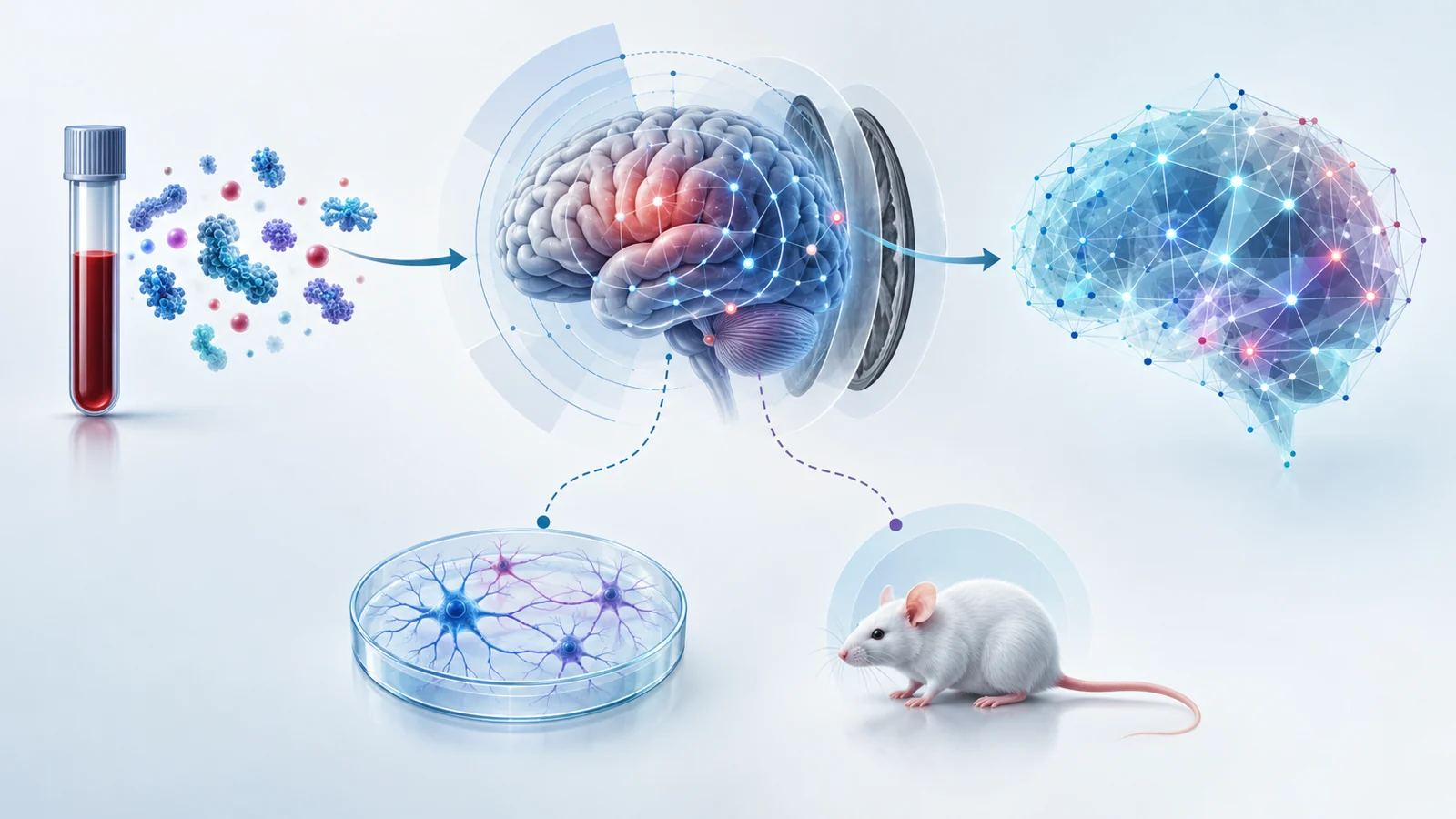
Parkinson’s disease is often discussed through its symptoms, but researchers also want to understand how molecular changes relate to altered brain function. A planned ClinicalTrials.gov study will examine whether blood-based measurements, brain imaging, clinical assessments, and laboratory experiments can be brought together to study that relationship.
The study focuses on alpha-synuclein, a protein associated with abnormal accumulation in Parkinson’s, and its possible interaction with tau. It is designed as a single-center, cross-sectional, nonrandomized interventional study with an estimated enrollment of 160 people. Its primary purpose is basic science, not testing a treatment. The supplied record lists the study as not yet recruiting, with estimated start and completion dates, and reports no posted results.
One study six ways to compare Parkinson’s biology
The human part of the study is organized around six groups. Some participants have Parkinson’s disease and some do not. Some have idiopathic REM sleep behavior disorder, a condition included here as part of the broader Parkinson’s spectrum under investigation. Within these clinical categories, the study separates people who carry a GBA1 mutation from those who do not. Healthy controls are included as another comparison group.
This arrangement lets investigators ask related questions at the same time. Do people with Parkinson’s show different molecular or brain-network patterns from people without the disease? Are similar patterns present in participants with idiopathic REM sleep behavior disorder? Do GBA1 carriers show a different relationship between protein-related measurements, brain connectivity, and clinical features than noncarriers?
The purpose is not to treat the six groups as six stages in a proven sequence. They are planned comparison groups. By separating genetic background from clinical presentation, the researchers hope to distinguish patterns associated with Parkinson’s, patterns associated with earlier or less established disease manifestations, and patterns associated with GBA1 status.
What blood samples and MRI can each reveal
Every participant is expected to undergo clinical and neuropsychological assessment alongside blood collection and multimodal MRI. These measurements answer different kinds of questions.
The blood analyses are intended to describe several biological processes. Planned measurements include total alpha-synuclein and a modified form called 129P-alpha-synuclein in neuronal-derived extracellular vesicles, as well as total tau and phosphorylated tau. The researchers also plan to measure glucocerebrosidase activity in peripheral blood cells, circulating cell-free mitochondrial DNA, and proteins linked to synaptic integrity such as SNAP25, STX1A, VAMP2, and neurogranin. Neurofilament light chain is another planned measure related to neurodegeneration.
A blood result would therefore provide a molecular snapshot, not a direct recording of protein movement through the brain. It could help show whether biological profiles differ between the study groups and whether those profiles relate to clinical or imaging observations.
MRI supplies a different view. Structural sequences can describe brain anatomy, diffusion-weighted imaging can provide information about white-matter tract integrity, and resting-state functional MRI can be used to examine how brain regions are connected when the participant is not performing a task. The study plans to extract network measures such as global and nodal efficiency, participation coefficient, and white-matter connectivity indexes.
Clinical and cognitive assessments give those measurements a human context. They can be compared with motor, nonmotor, and cognitive manifestations recorded during the same baseline evaluation. The value of the design comes from placing the molecular, network, and clinical observations side by side rather than treating any one measurement as a complete explanation.
Why the study includes cells and mice
Human group comparisons can show that characteristics occur together, but they are limited in what they can reveal about the biological response to a specific exposure. The study therefore adds controlled experimental work.
A small subset of 10 participants is planned to undergo a 3–4 millimeter skin biopsy under local anesthesia. Cells from these samples will be used to generate participant-specific induced pluripotent stem cell-derived dopamine-producing neurons. Researchers plan to investigate neuronal and synaptic responses in those cells in relation to each person’s biomarker profile and genetic background. The cell experiments will also expose neurons to alpha-synuclein and to a combination of alpha-synuclein and tau.
The animal work creates another controlled setting. GBA-associated Parkinson’s mouse models and wild-type mice are planned for behavioral testing, MRI and PET imaging, and postmortem assessment of synaptic, axonal, and mitochondrial pathology. Some mice will receive saline, some alpha-synuclein, and some combined alpha-synuclein and tau fibrils. These manipulations are intended to help researchers examine how defined experimental conditions relate to behavior, imaging changes, and tissue-level findings.
The same preclinical program includes validation of a new alpha-synuclein PET tracer called [¹⁸F]Syntacasyn. That work is planned validation in animal models. It should not be read as evidence that a validated human PET test already exists.
How a baseline snapshot becomes a computational model
The investigators plan to integrate the human and animal data with statistical and computational methods. One goal is to identify network hubs that appear particularly vulnerable when molecular, imaging, and clinical information are considered together. Another is to generate subject-specific “virtual brain” models that represent possible patterns of alpha-synuclein pathology propagation.
Such a model could help organize observations that are difficult to interpret separately. For example, a blood profile, a pattern of white-matter integrity, and altered functional connectivity might fit a common biological hypothesis more closely when analyzed together than when each result is viewed in isolation.
The design also sets a clear limit on what the first human measurements can establish. The listed human outcomes are measured at baseline, and the imaging procedure describes a single MRI acquisition. Comparisons at that point can identify relationships and support hypotheses, but they do not show the temporal order in which protein accumulation, network changes, and symptoms develop within an individual. They also do not demonstrate causal protein spread, predict how a particular person’s disease will progress, or establish diagnostic accuracy or clinical benefit.
That limitation is central to interpreting the study. The computational models are intended to generate and test biologically informed explanations, not to turn one cross-sectional assessment into a proven time-lapse of disease.
What this could change for understanding individual Parkinson’s disease
If the relationships are reproducible and the computational models can be validated, this approach could give researchers a more biologically specific way to understand why Parkinson’s disease differs between people. A person’s molecular profile might eventually be considered alongside patterns of brain-network vulnerability, rather than relying on clinical presentation alone.
The potential value is especially relevant to GBA1 backgrounds, where the study is designed to examine how genetic status relates to alpha-synuclein, tau, mitochondrial and synaptic measures, and brain connectivity. Over time, consistent relationships could support more precise biomarker research and help define biologically meaningful participant groups for future studies. They would not, by themselves, guarantee an individual prediction tool or a treatment benefit. Those possibilities depend on findings from this and later validation work.
Key Sources
- Primary source: Understanding Alpha-Synuclein Spread in Parkinson's Disease Through Blood Biomarkers and Neuroimaging
Government or Regulator · Clinical Trial · 2026-03-16
Keep exploring