
Research Signal – Source date 2026-02-10
A cell has a recycling system that can protect it from damage. But under some conditions, the same system may help a damaged cell remain active and continue sending disruptive signals to its surroundings. That apparent contradiction is the central problem addressed by a review published on February 10, 2026, in Redox Biology.
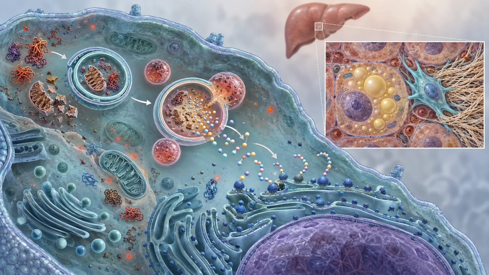
The review proposes a threshold model for the relationship between autophagy and cellular senescence. Autophagy is the process by which a cell encloses damaged proteins and organelles, delivers them to lysosomes for breakdown, and reuses the resulting materials. Senescence is a stress-associated state in which a cell stops dividing but can remain metabolically active. The model asks whether the recycling system is helping a cell stay functional or helping an already dysfunctional cell sustain its activity.
The proposal is explanatory rather than a ready-made clinical test. The proposed threshold should not be treated as a validated numerical cutoff or a clinical test that assigns every cell, organ, or patient to a universal before-or-after state. Its value is in showing why the same biological process could point toward different treatment strategies at different stages of damage.
A recycling system with two jobs
Under relatively stable conditions, basal autophagy acts as intracellular quality control. It removes protein aggregates, lipofuscin deposits, and malfunctioning organelles before they can further disturb the cell. Mitophagy, the selective removal of damaged mitochondria, is especially important in the review’s account because poorly functioning mitochondria can produce excess reactive oxygen species, or ROS. By clearing them, the cell can limit oxidative stress and preserve its ability to function.
This protective role helps explain why reduced autophagy is often associated with the conditions that precede senescence. A cell is exposed to many forms of stress at once, not just one. Mitochondrial ROS can combine with oxidative stress in the endoplasmic reticulum, where proteins are folded, and with hydrogen peroxide released by poorly cleared peroxisomes. The combined burden can exceed the cell’s ability to neutralize or remove damage.
Once the degradative system can no longer keep up, the cell may move toward a durable growth arrest. The review describes a temporary reduction in autophagy during this transition, alongside stabilization of p53 and activation of pathways involving p21 and p16. These signals help enforce the decision to stop dividing. The cell has crossed a functional threshold because interacting damage processes have overwhelmed its quality-control capacity, not because it has reached a particular age or a universal molecular reading.
How recycling supports established senescent cells
The model’s unusual feature is what happens after senescence is established. The reduction in autophagy is described as temporary. Recycling activity can resume, but its role has changed. Instead of primarily preventing the buildup of damage in a functioning cell, it can help a nondividing senescent cell meet the unusually high material and energy demands of continued secretion.
Senescent cells can produce a senescence-associated secretory phenotype, commonly abbreviated as SASP. In plain language, this means that they release a mixture of inflammatory cytokines, growth factors, and tissue-remodeling enzymes. The SASP can alter nearby cells and disturb tissue organization even though the senescent cell itself no longer proliferates.
The review links this secretory workload to a specialized arrangement called the TOR–autophagy spatial coupling compartment, or TASCC. In this arrangement, autolysosomes, where cellular material is broken down, are positioned near active mTORC1 growth signaling close to the trans-Golgi network. The arrangement matters because it puts recycling and protein production in close proximity.
The material flow is direct. Autophagy breaks down intracellular proteins and other components into smaller products, including amino acids such as arginine and leucine. Rag GTPases sense these amino acids near the lysosomal membrane and help reactivate mTORC1 in the same local compartment. The recycled building blocks can then support the synthesis of proteins that are exported from the cell, including SASP components such as IL-6 and IL-8.
A cell that has stopped dividing therefore still has a substantial production problem to solve. Spatially coupling recycling with growth signaling gives it a way to keep supplying that production line from its own contents, even when the wider environment is stressful. The threshold model describes this as a functional inversion: recycling that once helped restrain damage is repurposed to support the survival and inflammatory output of an established senescent cell.
One liver, different cellular problems
The review’s liver example shows why a treatment cannot be chosen only by looking at an organ’s overall condition. In early metabolic liver disease, hepatocytes may receive more free fatty acids than they can safely process. Lipid droplets expand, and lipophagy—the autophagic clearance of those droplets—can become inadequate. Impaired lysosomal acid lipase activity is described as one factor that limits lipid breakdown. Retained lipids can undergo oxidative damage, producing reactive aldehydes such as 4-hydroxynonenal that further stress mitochondria and push hepatocytes toward senescence.
The neighboring stellate cells can face a different problem after that transition. Senescent hepatocytes may release signals including transforming growth factor beta and Hedgehog ligands. These signals can activate previously quiescent hepatic stellate cells, turning them into collagen-producing myofibroblasts. In those activated stellate cells, autophagy may be increased to supply the energetic and biosynthetic demands of making extracellular matrix—the material that accumulates during scarring.
The same tissue can thus contain two relevant cellular situations. In hepatocytes, inadequate lipid recycling may help initiate damage and senescence. In activated stellate cells, recycling may support the production of scar tissue after the local injury has developed. A whole-organ intervention that simply raises or lowers autophagy could affect both populations at once, despite their different needs.
This is also why the model does not turn “autophagy” into a universal good or bad label. The useful question becomes more specific: which cell type is being changed, what stage of damage has it reached, and which activity is the treatment meant to interrupt or preserve?
Clinical research is ahead of diagnosis
The review connects its model to a reported portfolio of 13 registered studies involving autophagy-related interventions in aging- and senescence-related settings. The examples include approaches intended to reinforce cytoprotective processes, such as spermidine, fasting-mimicking diets, time-restricted eating, NAD+ precursors, exercise, and nutritional interventions. The review also includes two supplementary regenerative studies that address tissue repair and cellular replenishment alongside autophagy-centered strategies.
The portfolio is presented as clinical research activity organized around two broad aims: strengthening cellular maintenance before severe dysfunction, and disrupting survival pathways after senescence has become established. That inventory shows that the field is testing ideas across cancer, neurodegeneration, metabolic liver disease, fibrosis, immune aging, and tissue repair. It does not by itself show that clinicians can already determine a patient’s position relative to the proposed threshold or improve outcomes by assigning treatment according to it.
The authors therefore place substantial weight on functional biomarkers and patient stratification. Candidate measurements discussed in the review include circulating lncRNA TERRA, growth differentiation factor 15, and imaging-based assessments of mitophagy flux. These are candidates for defining where an individual or tissue may sit along a senescence trajectory, not established universal gauges of a patient’s treatment state.
A practical test of the model would require more than measuring whether autophagy is present. It would require identifying which cells are using it, whether recycling is restoring quality control or feeding a secretory program, and whether a proposed intervention reaches the relevant population without worsening another one. The difference between activating autophagy for prevention and inhibiting it, or using senolysis, for established disease depends on answering those questions reliably.
What this could mean for treating age-related disease
The longer-term possibility is a more selective form of treatment: preserving recycling in cells that can still recover while disrupting the survival or inflammatory activity of harmful senescent cells. Patients with tissue damage could benefit from better-targeted interventions if the threshold model proves useful for predicting responses, relevant cell populations can be identified, and the approaches are shown to be safe and effective.
From the review’s February 2026 perspective, that possibility remains conditional. The mechanistic sequence gives researchers a way to connect cellular quality control, inflammatory secretion, and disease progression, but clinical use would depend on validated functional biomarkers and treatment strategies that respect differences between neighboring cells. The promise is not a general instruction to boost or block autophagy. It is the prospect of matching the intervention to the cell, the stage, and the job that recycling is performing at that moment.
Key Sources
- Primary source: The autophagy-senescence axis as a threshold model of aging and therapeutic targeting.
DOI or Journal Page · Review or Meta-Analysis · 2026-02-10
Keep exploring