
Research Signal – Source date 2026-02-01
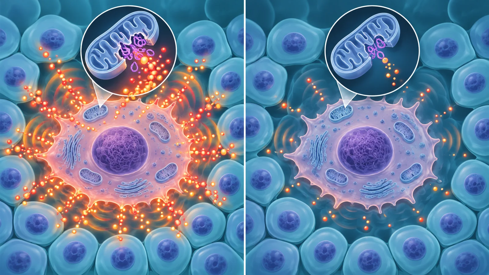
Aging research often treats cellular senescence as a problem to remove. But senescence has more than one feature. A senescent cell has entered a durable state in which it no longer divides, yet it may continue releasing molecules that alter the tissue around it. Those inflammatory messages are commonly grouped under the term senescence-associated secretory phenotype, or SASP.
The distinction matters because an intervention could reduce a cell’s inflammatory output without removing the cell or reversing its cell-cycle arrest. Would that help an aging organ, or merely change one measurable feature of senescence?
A report published February 1, 2026, describes research presented at the Sixth Annual Research Symposium of the Midwest Aging Consortium, held at Mayo Clinic in Rochester, Minnesota, from April 23 to 25, 2025. The meeting covered a broad range of aging research. One connected direction examined how senescent-cell behavior is shaped by mitochondria, tissue location, immune surveillance, and measurable blood markers.
Why stopping division is not the whole story
Mitochondria are structures inside cells that help manage energy and other metabolic processes. In the symposium report, researchers described evidence that damaged or stressed mitochondria can also influence the inflammatory program of senescent cells.
One set of experiments focused on BAX and BAK, proteins that can create openings in mitochondrial membranes. In senescent cells, leakage of mitochondrial DNA into the surrounding cell fluid activated inflammatory pathways, including cGAS-STING and NF-κB. Mitochondrial RNA leakage activated another set of innate immune sensors, MDA5 and RIG-I, with downstream signaling through MAVS. These processes helped drive the SASP, while the cells remained in their durable non-dividing state.
The reported interventions separated those features experimentally. Deleting BAX and BAK reduced mitochondrial DNA and RNA leakage and weakened inflammatory signaling without removing the cell-cycle arrest. In aged mice, the same genetic change was associated with lower inflammation. A pharmacological BAX inhibitor was also reported to improve healthspan measures and reduce senescence markers across several organs in mice.
A second presentation examined a different route into the same problem. Mitochondrial metabolites can affect gene activity by influencing epigenetic processes, which help determine whether particular genes are available for expression. Clearing mitochondria from human fibroblasts reduced histone acetylation at key SASP gene promoters. Inhibiting the mitochondrial citrate transporter SLC25A1 in senescent cells also reduced histone acetylation and SASP-factor expression without changing cell-cycle arrest.
The citrate-related pathway therefore offered a different way to reduce inflammatory output. In aged mice, inhibiting SLC25A1 was reported to reduce systemic inflammation and frailty while improving muscle and heart physiology. These findings make the separation between cell-cycle arrest and inflammatory secretion experimentally useful. They do not show that either approach is a safe or effective treatment for preserving function in older people, and they do not establish that every beneficial or protective function of a senescent cell would remain intact.
Location changes the question
A senescent cell does not affect tissue in isolation. Its position, neighboring cells, and local signals may determine whether its presence contributes to repair, inflammation, or disease. The symposium report described spatial transcriptomics as one way to examine that context. The method measures patterns of gene activity while retaining information about where those patterns occur in a tissue.
In lung samples, Jhonny Rodriguez Lopez and colleagues used spatial patterns, single-cell RNA sequencing profiles, deep learning, and Bayesian modeling to identify regions associated with senescence in healthy aging and idiopathic pulmonary fibrosis. The analysis considered three anatomical areas: the lung parenchyma, peri-bronchial regions, and subpleural areas. Senescence-associated genes were enriched in particular locations rather than appearing as a uniform signal throughout the lung. In healthy-aged specimens, Cdkn1a-associated activity was especially prominent around the bronchi, while in fibrotic lungs it was more pronounced in upper lung regions.
The analysis also linked specific spatial patterns with cell types associated with fibrosis, including CTHRC1-positive fibroblasts and aberrant basaloid epithelial cells. The value of this approach is not simply that it produces a more detailed map. It can help researchers ask whether senescence-associated activity is concentrated near the cells and structures most relevant to tissue dysfunction.
Immune cells add another layer. Natural killer cells can help identify and respond to abnormal cells, so changes in their activity could affect how senescent cells accumulate. In a mouse model of bleomycin-induced lung injury, depleting natural killer cells led to more senescent cells and greater fibrosis. Separate analyses of human lung tissue from people with idiopathic pulmonary fibrosis and from healthy aged donors found that lung natural killer cells in fibrosis had lower expression of cytotoxicity-related genes and greater expression of genes associated with an ILC1-like state, including PD-1.
The human tissue findings also showed more ILC1-like natural killer cells in fibrotic lungs and lymph nodes. Fibroblasts with senescence features expressed the corresponding ligand, PD-L1. In cell-culture experiments, conditioned medium from fibrotic lung fibroblasts was more effective at shifting natural killer cells toward an ILC1-like phenotype than medium from young or aged healthy donors.
These observations come from different types of evidence. The spatial and immune findings in human specimens describe associations in diseased tissue, while the natural-killer-cell depletion experiment in mice tests a perturbation in a model. Together they show why location and immune behavior matter when studying senescence, but they do not constitute one treatment pathway tested from intracellular mechanism through human organ recovery.
A blood marker can track biology without proving benefit
The report also described IL-23R as a candidate circulating marker of aging and senescence. In aged mice, IL-23R was higher in plasma than in young mice, and senescent mouse fibroblasts produced more of the protein. Its expression also increased with age in several tissues, including the kidney and liver.
The response varied depending on what was measured. Venetoclax reduced plasma IL-23R in male and female mice. Tissue measurements differed by intervention and by sex. The aged kidney appeared to be a likely source of some age-related circulating IL-23R in the reported experiments. In plasma samples from Mayo Clinic Biobank participants between 20 and 82 years old, IL-23R also increased with age in both males and females.
That makes IL-23R useful as a concrete example of the difference between a biological response and a functional outcome. A treatment that lowers a blood marker may have changed one pathway associated with senescence. It does not automatically show that the treatment improved strength, mobility, organ performance, or health in a person. The supplied findings do not establish IL-23R as a validated diagnostic test or as a surrogate endpoint for better health.
The same caution applies to the broader evidence. The report brings together cultured-cell experiments, genetic and pharmacological interventions in mice, and analyses of human tissue. Those studies illuminate different parts of aging biology. They should not be read as evidence that a BAX-targeted strategy, an SLC25A1 inhibitor, or any other senescence-focused intervention has already demonstrated human benefit.
What quieter senescent cells could mean for healthy aging
The most practical possibility raised by this research is not necessarily the wholesale removal of senescent cells. It is the prospect of reducing damaging inflammatory output while preserving the cells’ non-dividing state, provided that the relevant mechanisms translate to people and treatment can reach the intended tissues without unacceptable effects.
The mouse findings involving mitochondrial pathways point toward a conditional clinical idea. Lowering inflammatory signaling might eventually help protect the environments around aging muscle and heart tissue, potentially supporting everyday physical function in older adults. That possibility remains dependent on several links in the chain: the molecular mechanism must operate similarly in humans, a treatment must act in the right organs, and the benefits must outweigh toxicity or disruption of useful cellular roles.
For now, the symposium report supports a more precise question about senescence. Researchers do not have to choose only between leaving senescent cells untouched and eliminating them. They can also investigate which signals make those cells harmful, where those signals matter most, and whether changing them leads to meaningful improvements in how aging organs work. The important endpoint would not be a quieter blood marker alone, but better function in daily life.
Key Sources
- Primary source: The Sixth Annual Symposium of the Midwest Aging Consortium.
DOI or Journal Page · Peer-Reviewed Paper · 2026-02-01
Keep exploring