
Research Signal – Source date 2025-10-14
The heart and blood vessels do not age through one isolated process. A review published on October 14, 2025, connects chronic low-grade inflammation, cellular senescence and changes to the extracellular matrix, the supporting material that surrounds cells. Together, these processes can alter how heart muscle and arteries stretch, contract, repair themselves and withstand stress.
That biology also creates a difficult therapeutic question. If senescent cells release inflammatory signals and enzymes that damage surrounding tissue, removing them may seem helpful. Yet senescence can sometimes take part in repair or limit excessive scarring. The outcome depends on which cells are involved, where they are located and what the tissue is trying to do at that stage of disease.
The tissue around cells is part of the cardiovascular system
The extracellular matrix is not passive packing material. It is a dynamic network of proteins, proteoglycans, polysaccharides and signaling molecules that gives tissue structure while communicating with the cells inside it. In the heart and vessels, interstitial matrix components such as collagen, fibronectin and elastin help organize tissue and contribute to its mechanical properties. Basement membranes provide another specialized support layer around cells.
The matrix is continually adjusted. Cells produce new components, while enzymes break down older material so the tissue can respond to injury and changing mechanical demands. This turnover is useful when it is controlled. Persistent inflammation, hypoxia, biomechanical stress or excessive neurohormonal activation can push the process toward maladaptive fibrosis and organ dysfunction.
A remodeled matrix can become too abundant, too stiff or poorly organized. In other settings, its protective structure can be degraded faster than it is rebuilt. These are different physical failures, but both can interfere with cardiovascular function.
Inflammation gives aging cells a larger reach
Inflammaging refers to a persistent, moderate inflammatory state associated with aging. It differs from the short-lived response to an infection or an acute injury. The review describes elevated inflammatory mediators such as TNF-α, IL-1β, IL-6 and C-reactive protein as part of this broader environment, which is also linked with mitochondrial dysfunction and changes in immune regulation.
Cellular senescence adds another layer. A senescent cell has permanently left the cell cycle and often becomes resistant to programmed cell death, but senescence is not a single uniform condition. Its features vary according to cell type and surrounding conditions. Some senescent cells alter their metabolism, produce more reactive oxygen species and release a collection of substances known as the senescence-associated secretory phenotype, or SASP.
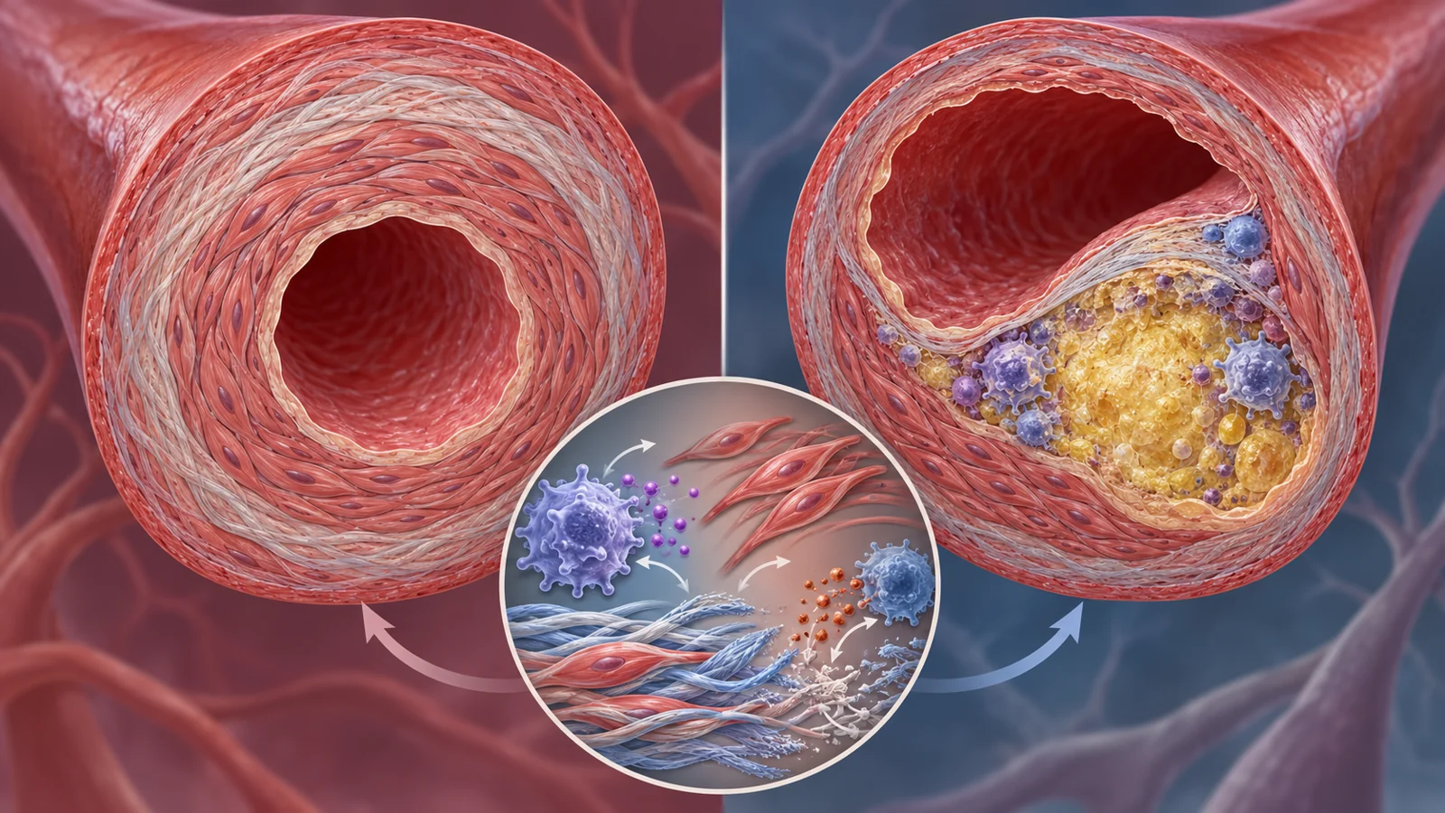
The SASP can include cytokines, chemokines, growth factors, microRNAs, damage-associated signals and proteolytic enzymes. Matrix metalloproteinases are especially important here because they can break down extracellular matrix proteins. Other SASP components can recruit and activate immune cells, influence neighboring cells or impair the function of nearby tissue-supporting cells.
Inflammation can also increase matrix production by activating fibroblasts, while simultaneously increasing matrix-degrading enzymes. Macrophages participate in this process by secreting MMPs and by ingesting matrix components. Fragments released during matrix breakdown can then influence macrophage behavior and movement. The matrix is therefore both a target of inflammatory activity and a source of new signals that can sustain it.
A stiff artery and a fragile plaque are different failures
In hypertension, the dominant structural problem described by the review is excessive vascular remodeling. Sustained renin-angiotensin-aldosterone system activity, inflammation and oxidative stress can encourage endothelial and smooth-muscle cells to take on myofibroblast-like behavior. Excess extracellular matrix deposition thickens the vessel wall, reduces elasticity and increases vascular resistance.
Changes in small resistance arteries and large conduit arteries can reinforce one another. A vessel that has become less flexible does not respond to blood flow in the same way as a healthy vessel, while increased resistance adds further stress to the circulation. The result is a physical environment that can help maintain hypertension and impair tissue perfusion.
Atherosclerotic plaque involves a different structural danger. In an enlarging plaque, inflammatory cells such as macrophages and T lymphocytes release cytokines and proteolytic enzymes, including MMPs that can degrade collagen in the fibrous cap. The cap is part of the structure separating the plaque’s lipid-rich core from the blood. If it becomes thin and weakened, the plaque may be more vulnerable to rupture.
The review discusses thin-cap fibroatheromas as plaques with caps less than 65 micrometers thick, large lipid cores and inflammation. It also describes microhemorrhages and fragile new vessels within plaques as features that can accelerate plaque progression. Advanced ultrasound and MRI are being developed to assess structural and inflammatory features of plaque vulnerability, but the supplied evidence does not turn those approaches into a general prediction of individual risk.
The same broad network of inflammation, immune activity and matrix turnover can therefore produce excessive deposition in one setting and damaging structural loss in another. Understanding the physical change matters more than treating remodeling as a single process that always means too much tissue.
Why clearing senescent cells can be the wrong move
The apparent contradiction becomes clearer after myocardial infarction. Cardiac fibroblasts are central producers and organizers of the heart’s extracellular matrix. The review reports that activation of the p53/p21 pathway can induce fibroblast senescence after injury, and that this may limit fibrosis through senescence-associated factors such as CCN1.
That example does not make senescence beneficial in every situation. It shows why cell removal cannot be treated as a universal repair strategy. Senescent cells can contribute to wound healing, tissue remodeling and fibrosis under tightly regulated conditions, while their secretory activity can become harmful when the cells accumulate or remain active in an unfavorable environment.
Senolytics are drugs designed to selectively eliminate senescent cells. The review discusses agents including dasatinib, quercetin, fisetin and navitoclax, which act on anti-apoptotic pathways that help senescent cells survive. Preclinical studies have suggested potential benefits in several age-related conditions, and the cells may reaccumulate slowly enough for intermittent treatment to be considered.
But the cardiovascular evidence is not a simple success story. The review also reports conflicting results and adverse findings in animal models, including worsening cardiac dysfunction, destabilization of atherosclerotic plaques and increased mortality. These findings do not establish that senolytics are unsafe in every human cardiovascular context. They do establish why the relevant cell population, tissue location and phase of repair matter before a broad cell-clearing approach could be trusted.
Related treatments test different ideas
Several therapeutic approaches discussed in the review should not be treated as interchangeable. Human evidence for statins, for example, concerns an established treatment class with effects that extend beyond cholesterol lowering. In the JUPITER trial summary, 17,802 apparently healthy adults with LDL cholesterol below 130 mg/dL and high-sensitivity CRP of at least 2 mg/L were assigned to rosuvastatin or placebo. Rosuvastatin reduced LDL cholesterol by 50%, CRP by 37% and the primary endpoint by 44%.
The reductions in LDL cholesterol and CRP document lipid lowering and a fall in an inflammatory marker; the trial summary does not isolate which effects account for the clinical benefit. They do not demonstrate that rosuvastatin clears senescent cells, and they do not isolate extracellular matrix remodeling as the reason for the clinical result. A human result with one intervention cannot be transferred automatically to a different mechanism.
The review also describes a treatment experiment in OVE26 mice with diabetic cardiomyopathy. Mice received the proteasome inhibitor MG-132 at 10 micrograms per kilogram per day for three months. The reported improvements in cardiac function and remodeling were associated with lower oxidative stress and inflammation, increased Nrf2 activity and reduced NF-κB signaling. This is an experimental animal finding, not evidence that the same treatment is effective or safe in people.
Nrf2-related strategies face questions about bioavailability, toxicity and the risks of sustained activation. Matrix-directed treatments raise a different problem. MMPs help repair and maintain tissue as well as contributing to pathological remodeling, so broad inhibition could interfere with normal biology. The review presents selective inhibition of relevant enzyme isoforms or localized delivery to affected cardiac tissue as possible ways to limit adverse remodeling while preserving useful matrix turnover.
The same caution applies to RNA-based drugs, anti-inflammatory biologics and other emerging approaches. A therapy that changes inflammatory signaling, oxidative stress, senescent-cell survival or matrix enzymes is acting on a different part of the system. Evidence for one does not validate all the others, and none of these discussions establishes that cardiovascular aging has already been reversed clinically.
What targeted tissue repair could mean for aging hearts
The practical promise lies in controlling harmful remodeling without disabling the repair processes that keep cardiovascular tissue functional. For an older person, that could eventually mean preserving the flexibility of vessel walls, limiting scar-driven loss of cardiac performance or maintaining the structural integrity that helps a plaque remain stable.
Selective or localized treatment is one possible route to that outcome. A therapy delivered to the relevant tissue, aimed at a specific matrix-degrading enzyme or harmful inflammatory pathway, could be more useful than indiscriminate removal of every senescent cell. That possibility remains conditional on demonstrating safe, effective targeting in the right patients and disease settings.
If those constraints can be met, understanding the conversation between aging cells and the extracellular matrix could lead to cardiovascular treatments that preserve function rather than simply suppressing one risk factor. The goal would be an aging heart that retains more of its mechanical resilience and arteries that remain more capable of adapting to the demands placed on them.
Key Sources
- Primary source: Inflammaging and Senescence-Driven Extracellular Matrix Remodeling in Age-Associated Cardiovascular Disease.
DOI or Journal Page · Review or Meta-Analysis · 2025-10-14
Keep exploring