
Evidence date · 2025-09-16 · Research note 1 of 3
1 2 3
See current Track status →
The sleep disorder raises a question about what comes next
Some people with isolated REM sleep behavior disorder act out their dreams because the usual muscle suppression of REM sleep is lost. They may move, kick, or vocalize while dreaming. The condition is important to researchers because it can appear years before a neurodegenerative disorder such as Parkinson disease or dementia with Lewy bodies becomes clinically apparent.
The difficult question is not simply whether people with this sleep disorder differ from healthy adults. It is whether a measurable brain change can help distinguish which disease trajectory may follow. A study published on September 16, 2025, examined that question with MRI scans from five contributing centers or cohort sources. Its main finding connected a lower left-sided MRI index with later Parkinson diagnoses. The association is scientifically useful, but it does not turn one scan into a personal prediction.
What the MRI index is trying to measure
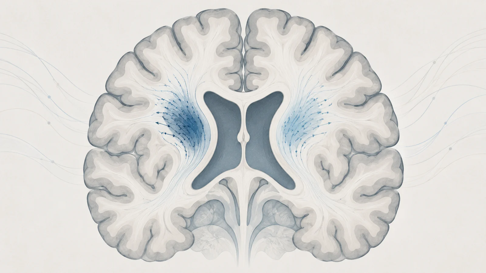
The study used diffusion tensor imaging along the perivascular space, shortened to DTI-ALPS. In plain terms, this is an MRI-derived index that compares how water appears to move in selected directions near the brain’s fluid-filled ventricles. The researchers used small regions in nearby white matter on each side of the brain and calculated the index from directional diffusivity measurements.
DTI-ALPS has been proposed as an indirect proxy for the glymphatic system, a set of fluid-related pathways involved in moving interstitial waste through the brain. That proposed relationship gives the measurement biological interest, especially because abnormal protein accumulation and impaired waste handling are relevant research questions in neurodegenerative disease. But the index itself measures regional water diffusivity. It does not directly measure protein removal, and it does not provide a whole-brain reading of waste clearance.
The investigators also examined conventional and free-water-related diffusion measures from the same regions. Those measures did not show significant differences in the reported comparisons between patients and controls or between converters and nonconverters. This made the result more specific to the DTI-ALPS calculation within this study, while leaving open what biological process accounts for the association.
How the five-center comparison was built
The final analysis included 250 people with isolated REM sleep behavior disorder and 178 healthy controls. The sleep disorder had been confirmed with video polysomnography, which documents REM sleep without the expected muscle atonia and helps exclude alternative explanations for nighttime movements or vocalizations. Participants had both structural and diffusion-weighted brain MRI.
Because the scans came from different centers and scanners, the researchers used a harmonization procedure to account for acquisition differences. They also performed image-quality checks, including checking whether the measurement regions were correctly placed. Some participants were excluded after processing failures, mask misregistration, or extreme values. These steps do not remove every source of variation, but they make a multicenter comparison more interpretable.
The group difference was modest. The average left DTI-ALPS index was 1.29 in the isolated REM sleep behavior disorder group and 1.32 in controls, with a Cohen d of 0.17. The right-sided comparison was not statistically significant. The finding therefore was not a large, bilateral separation between two populations. It was a small difference concentrated on the left side.
The baseline scan came before the later diagnoses
The study’s longitudinal contribution came from following 224 of the patients with isolated REM sleep behavior disorder for an average of 6.1 years, with follow-up ranging from 1 to 16 years. During that period, 65 received a recorded conversion diagnosis. At the last follow-up, 159 had not received such a diagnosis. That latter category describes their status during the observed period; it is not a lifelong guarantee that disease will never develop.
The people who later converted had a lower left DTI-ALPS index at baseline than those who remained without a conversion diagnosis at the last assessment. The average was 1.25 among converters and 1.30 among nonconverters. The scan therefore preceded the outcome being studied, which gives the association a temporal order that a one-time group comparison cannot provide.
The outcome group was not uniform. It included 42 Parkinson disease diagnoses and 18 dementia-with-Lewy-bodies diagnoses. The detailed breakdown also listed four multiple system atrophy diagnoses and one Alzheimer disease diagnosis. Those counts matter because the study was asking whether the same MRI feature related equally to different paths, not merely whether it separated everyone who later received any diagnosis from everyone else.
The Parkinson association was clearer than the dementia association
In the reported models, a lower left DTI-ALPS index was associated with conversion to Parkinson disease. The association persisted when cognitive or motor scores were added separately. In the model that included cognitive score, each standard-deviation increase in the left index was associated with lower odds of Parkinson conversion, with an odds ratio of 0.68 and a 95% confidence interval of 0.46 to 0.99.
That number describes an adjusted association within this study’s model. It is not the probability that a particular person will develop Parkinson disease, and it does not mean that raising the index would protect someone from disease. The analysis also did not establish a threshold that could be used to classify an individual in clinical practice.
The corresponding analyses for dementia with Lewy bodies did not show a statistically significant association with the left index. That pattern is informative, but it is not the same as demonstrating that the scan can reliably tell future Parkinson disease apart from future dementia with Lewy bodies. A significant result for one outcome and a nonsignificant result for another can arise from differences in sample size, statistical uncertainty, or the underlying relationships. Reliable discrimination would require validation in additional patients and evidence that the measurement adds useful information beyond established clinical assessments.
What the result can and cannot explain
The study supports a connection between a lower left-sided DTI-ALPS index and later Parkinson diagnoses among people with confirmed isolated REM sleep behavior disorder. It does not establish that impaired glymphatic clearance causes Parkinson disease. Nor does it show that changing the MRI index would change disease risk.
The measurement is regional and indirect. It samples water diffusivity in selected periventricular white-matter regions rather than directly observing fluid clearance throughout the brain. The left-sided pattern may eventually prove biologically meaningful, but this study alone cannot settle why it appeared or whether it will reproduce across scanners, populations, and analysis methods.
The follow-up also remains incomplete. Only a subset of the cohort had converted by the last assessment, and the disease-specific groups were small, particularly for outcomes other than Parkinson disease and dementia with Lewy bodies. The people classified as nonconverters had simply not received a conversion diagnosis during the available observation window.
The clinical correlations were similarly limited. Weak pooled correlations between the index and cognitive scores did not remain significant after adjustment for age and sex or after correction for multiple comparisons. The marker should therefore not be presented as an established measure of cognitive impairment. It is better understood here as a candidate progression-related MRI measure whose meaning still needs to be tested.
What this could mean for early Parkinson treatment trials
A reproducible version of this finding could help researchers assemble groups of people with confirmed isolated REM sleep behavior disorder who share a more similar likely trajectory toward Parkinson disease. That would make early treatment trials more informative by reducing some of the variation in who is likely to develop the outcome being studied and when.
The practical benefit would be in trial design, not proof that any treatment already prevents disease. For the MRI index to support that role, later studies would need to reproduce the association in additional patients, clarify whether it adds information beyond clinical measures, and show that its predictions remain useful across centers and imaging protocols. As of September 2025, the study provides a longitudinal clue toward that capability, not an actionable forecast for an individual.
Key Sources
- Primary source: Association of DTI-ALPS Glymphatic Index With Differential Phenoconversion in Isolated REM Sleep Behavior Disorder: A Multi-Cohort MRI Study.
DOI or Journal Page · Peer-Reviewed Paper · 2025-09-16