
Evidence date · 2022-06-17 · Research note 1 of 3
1 2 3
See current Track status →
Chronic obstructive pulmonary disease, or COPD, is often described as a disease of accelerated lung aging. One reason is the growing evidence that some lung cells enter senescence: they stop dividing but remain biologically active. These cells can continue releasing molecules that influence nearby tissue, even though they are no longer contributing normally to renewal.
A review published on June 17, 2022, titled “Senescence: Pathogenic Driver in Chronic Obstructive Pulmonary Disease,” brings together findings from patient lung tissue, cultured cells, animal models and selected drug studies. Its central question is not simply whether senescent cells can be detected in COPD. It is whether their persistence could help explain inflammation, failed repair and the structural changes that make the disease progressive.
The cells remain alive, but their role changes
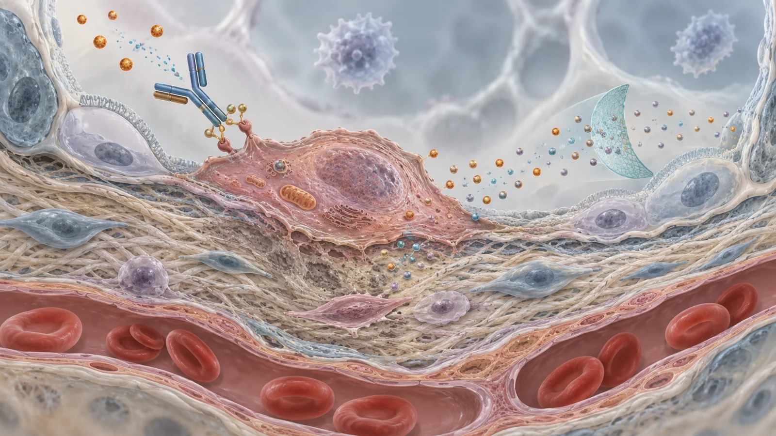
Cellular senescence is a persistent growth arrest accompanied by changes in cell shape and the release of extracellular factors. These secretions are commonly grouped under the term senescence-associated secretory phenotype, or SASP. They can include cytokines, growth factors and proteases, along with other cellular material that may affect neighboring cells.
No single marker identifies every senescent cell. Researchers commonly combine indicators such as the cell-cycle inhibitors p16 and p21, senescence-associated beta-galactosidase activity, altered telomere length, DNA-damage markers, lipofuscin accumulation and measurements of proliferative capacity. The need for several indicators matters because senescent cells vary by cell type and by the stress that produced them. SASP factors are also not exclusive to senescence; they can appear in other inflammatory settings.
The review describes higher levels of senescence-associated markers in several lung cell populations from people with COPD or emphysema. These include type II pneumocytes, endothelial cells, pulmonary artery smooth muscle cells, fibroblasts, small-airway epithelial cells and endothelial progenitor cells. Comparisons involved age-matched smokers, non-smokers or both, depending on the study. In some cases, marker levels or lower population-doubling capacity were associated with measures of airflow obstruction.
Those findings place senescence near important features of COPD, but they do not determine how much disease progression is caused by senescent cells. A marker can identify a stressed or altered cell without proving that the cell is driving the patient’s decline.
Smoke-related stress can push lung cells toward senescence
The proposed sequence begins with damaging exposure. Cigarette smoke can produce oxidative stress, DNA damage and mitochondrial dysfunction. Mitochondria are not only energy-producing structures; damaged mitochondria can accumulate when the systems that remove them, including pathways involving PINK1 and Parkin, work poorly. The resulting imbalance may increase reactive oxygen species and intensify cellular stress.
In cultured lung cells, cigarette smoke extract has been associated with senescence through pathways involving ATM, p53 and p21, as well as p16 and retinoblastoma-related signaling. Repeated exposure was important in experiments that produced an irreversible senescent state. Mouse lung tissue exposed to acute or chronic smoke also showed increased senescence-associated beta-galactosidase activity, p16, p21 or p53, and lipofuscin.
A smoke extract in a dish is not the same exposure as smoke entering a living lung. The concentration, timing and surrounding tissue environment differ, so cell-culture findings help test mechanisms without reproducing the whole disease. Even so, the experiments give a plausible connection between smoke-associated injury and the accumulation of damaged, persistently altered cells described in COPD.
Inflammation can then reinforce the cycle. Senescent cells may release IL-6, IL-8, MCP proteins, matrix metalloproteinases, TGF-beta and other mediators. They can also release vesicles, non-coding RNAs and mitochondrial material that influence neighboring cells. Chronic inflammatory stress, in turn, can promote senescence in additional cells. In COPD, reduced antioxidant defenses and elevated oxidative stress may make this feedback harder to resolve.
Failed replacement turns cellular stress into tissue damage
The lung is affected not only by inflammatory signals but also by the balance between cell loss and cell replacement. Alveolar epithelial and endothelial cells undergo apoptosis in emphysematous lungs, while the capacity to replace them can be limited. Increased p16 expression has been associated with lower expression of a proliferation marker in alveolar and vascular cells, connecting senescence-associated signaling with reduced renewal.
This creates a practical problem for damaged tissue. If cells die faster than they can be replaced, or if progenitor and repair cells stop dividing, the lung may lose part of its ability to restore its structure. Short telomeres limited epithelial recovery after smoke exposure in mice, while p21 deficiency promoted proliferation of some alveolar and airway cells in one model. Smoke exposure also inhibited normal fibroblast proliferation needed for repair before chronic exposure produced an irreversible senescent state.
The consequences may not be uniform. Extended smoke-extract exposure produced both senescent and non-senescent fibroblast phenotypes in one set of experiments. The senescent cells were discussed in relation to emphysema, while the non-senescent cells showed stronger profibrotic signaling. In other words, different responses to the same broad injury may contribute to different kinds of remodeling. Reduced regenerative capacity in airway basal progenitor cells has also been observed in COPD, although the cited evidence did not attribute that particular defect to senescence.
This is why the biological story is more complicated than “old cells accumulate and should be removed.” Senescence can have useful roles in development, tissue remodeling and the controlled response to certain stresses. Its effects depend on the cell, the trigger and the secretory pattern. A persistent harmful state in an adult lung is not interchangeable with every process that carries the name senescence.
Mouse results separate detection from causation
Animal experiments illustrate the difficulty of moving from association to intervention. In some smoke-exposure studies, removing or reducing p16-related activity lowered inflammatory cytokine expression, increased type II pneumocyte proliferation and protected mice from emphysema development. Other work found protection against inflammatory cell influx after acute smoke exposure but no improvement in lung-function decline or airspace enlargement during chronic exposure.
The review notes that differences in mouse models may contribute to these outcomes. The studies did not produce one uniform answer about what p16 controls, which cells matter most or which stage of disease is affected. A chronic-exposure result without protection against key structural or functional outcomes is especially important: changing a senescence-associated pathway is not automatically the same as preserving lung function.
The same caution applies to laboratory measurements. Higher p16, p21 or beta-galactosidase activity can support a senescence interpretation when combined with functional evidence, but no one indicator captures every relevant cell. COPD involves multiple interacting pathways, and p16-deficient mice did not show the same outcome across the reported experiments. Identifying a plausible driver and proving that it can be safely changed are separate scientific tasks.
Removing cells is different from changing their signals
The therapeutic ideas discussed in the review fall into more than one category. Senolytics aim to eliminate selected senescent cells. Other approaches try to prevent senescence, alter pathways that sustain it or reduce the SASP without removing the cells themselves. These strategies could have different risks because a treatment that clears cells may affect tissue composition, while a treatment that changes secretion may leave the underlying growth arrest in place.
Cardiac glycosides provide one experimental example. In cultured cells, ouabain and digoxin showed senolytic effects against p16-expressing human airway epithelial cells, and related experiments found elimination of senescence-induced lung fibroblasts. These observations suggest a possible way to test selective vulnerability, but they remain cell-based findings. They do not establish a safe COPD treatment or show that clearing such cells slows disease in people.
Rapamycin offers a different example. By inhibiting mTOR, it prevented or reduced senescence-associated changes and inflammatory SASP activity in mouse and lung-cell experiments. In smoke-exposed mice, the review describes reductions in inflammatory cells and measures of airspace destruction. These results are relevant to the possibility of modifying a pathway and its secretions, but they do not show that rapamycin produces the same effect in patients with COPD.
The review also discusses p38 MAPK inhibition. Acumapimod was associated with an improvement in forced expiratory volume in one second compared with placebo in clinical trials for severe acute COPD exacerbations. That lung-function finding is clinically relevant, but it does not by itself demonstrate that the benefit came from senescent-cell removal or senescence-specific signaling. A drug can influence inflammation or lung function through several mechanisms at once.
What this could change
For people living with COPD, the potential value of this line of research is practical: a treatment aimed at persistent damaging cell states might help preserve remaining lung function by reducing ongoing injury and protecting the capacity for repair. Reaching that benefit would require more than lowering a marker or changing an inflammatory measurement. Researchers would need to identify which cells and secreted signals sustain disease in patients, target them without disrupting useful senescence responses, and show a durable improvement in meaningful clinical outcomes.
The June 2022 review therefore presents senescence as a promising explanation and therapeutic direction, not a completed treatment breakthrough. If the links between damaged mitochondria, persistent growth arrest, inflammatory secretion and failed replacement can be separated clearly enough, future therapies could be designed around the part of the cycle that is both harmful and safely reversible. The patient-facing test would be preserved lung function and less ongoing damage, rather than a laboratory result alone.
Key Sources
- Primary source: Senescence: Pathogenic Driver in Chronic Obstructive Pulmonary Disease.
DOI or Journal Page · Review or Meta-Analysis · 2022-06-17