
Evidence date · 2022-03-29 · Research note 2 of 2
1 2
See current Track status →
Since the previous research note · Earlier expectation not tested
Track continuity
Since the previous research note: How nondividing cells can still affect kidney health
Previous expectation: A senescence-directed treatment during donor-kidney perfusion might improve the organ's resilience and function after transplantation while avoiding unacceptable damage to useful cells.
What this study adds: The March 2022 study characterized an engineered human kidney tubule cell model with senescence-like features and differential responses to senolytic drugs. It adds an experimental system for investigating cell susceptibility without testing donor-organ perfusion or graft outcomes.
What changed in this study
Kidney researchers face a difficult measurement problem when they study senolytics, a class of drugs intended to clear senescent cells. A senescent cell has entered a lasting growth arrest but may remain active in other ways, releasing signals that affect neighboring tissue. A drug that lowers the number of these cells could be useful, but a drug that simply kills cells broadly would not solve the same problem.
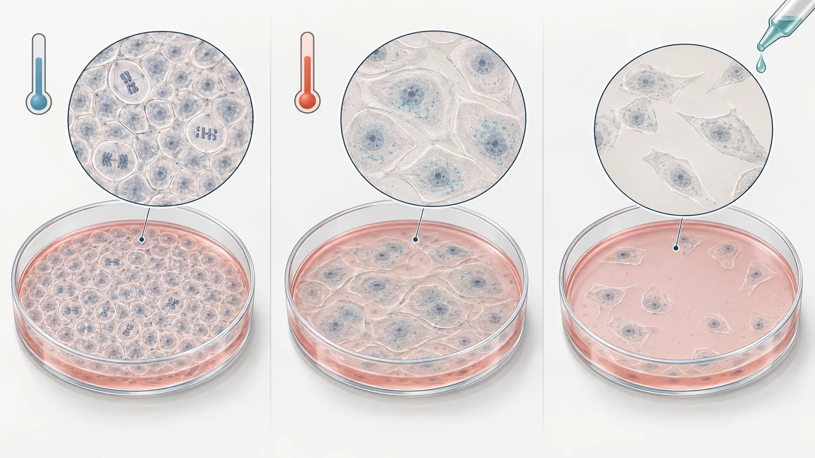
A March 29, 2022 study tested a way to make that comparison in a controlled human kidney-cell system. The researchers used an existing proximal tubule epithelial cell line and changed its culture temperature. At 33°C, the cells proliferated. At 37°C, they developed a more evident senescence-like state over time. The experiment therefore offered a way to ask which cells became more vulnerable to particular drugs, while keeping the comparison inside the same engineered cell model.
The work was laboratory research in cultured cells. It did not test a donor kidney, kidney function, fibrosis reversal, or treatment in people. Its contribution was narrower but useful: it characterized a controllable cell state and compared drug responses within that system.
Temperature changes the cells’ growth program
The cell line was conditionally immortalized, which means its ability to keep proliferating could be adjusted through an engineered temperature-sensitive mechanism. The system used a modified form of the SV40 large T antigen. At the permissive temperature of 33°C, that mechanism supported continued cell growth. Moving the culture to the non-permissive temperature of 37°C reduced the relevant SV40T activity and allowed growth-control pathways involving p53 and Rb to become more active.
The cells also expressed human telomerase, or hTERT. That helped maintain telomere length and prevented the model from being driven simply by the telomere-shortening process associated with replicative aging. This distinction matters. The experiment created a temperature-controlled growth arrest with senescence-like features; it was not a miniature version of every process that causes kidney cells to age naturally.
The researchers followed cells at days 0, 3, 6, 9, and 12 while keeping parallel cultures at 33°C or 37°C. Day 0 served as the non-senescent comparison, and the longer observations showed how the phenotype developed rather than treating a single measurement as decisive. By about nine days at 37°C, several findings had aligned strongly enough for the authors to select that time point for testing senolytics.
Several measurements build the senescence-like case
The evidence came from different kinds of measurements, and they did not all tell an identical story. At seven days, cells held at 37°C had substantially higher p21 RNA than cells at 33°C. P21 is associated with cell-cycle arrest, so this result fit the observation that the cells were no longer progressing through growth in the same way. Lamin B1 RNA moved in the opposite direction, decreasing at 37°C. Lamin B1 is a structural protein associated with the cell nucleus, and its loss is commonly used as one indicator of senescence-related change.
The protein measurements added detail rather than simply repeating the RNA results. P21 protein increased in the 37°C cultures during the maturation period. Total p53 showed a different pattern, with higher levels in the proliferating 33°C group and a decreasing trend in the 37°C group. Lamin B1 protein showed a downward trend, especially with longer culture, but this trend was not statistically significant. RNA expression and protein abundance therefore need to be kept separate; a change in one is not automatically proof of the same change in the other.
The cells also became positive for senescence-associated beta-galactosidase, usually called SA-β-gal. This staining reflects increased lysosomal activity often seen in senescent cells. At 37°C, beta-gal expression and activity rose during maturation, with the clearest staining appearing after the cells had spent several days under the non-permissive condition. The staining supported the other observations, but it was part of a group of measurements rather than a standalone definition of the cell state.
Together, growth arrest, p21 changes, reduced Lamin B1, and SA-β-gal activity gave the researchers a reason to describe the 37°C cultures as senescence-like. The wording is important. The model reproduced selected features of senescence in a human proximal tubule cell line, not every pathway or consequence of naturally accumulated senescent kidney tissue.
The signals outside the cell were uneven
Senescent cells matter partly because of what they release. Their senescence-associated secretory phenotype, or SASP, can include inflammatory and profibrotic factors. In kidney research, those extracellular signals are relevant because they may influence nearby cells and processes associated with fibrosis.
The study found that the secretory pattern depended on the molecule being measured. IL-6 secretion increased over time in both temperature conditions, but it was significantly higher in the 37°C cultures on days 3, 6, and 12. IL-8 also rose with time and showed a higher trend at 37°C. CTGF was slightly higher at the non-permissive temperature, although its levels rose and then moved back toward baseline during the observation period.
TNF-α provided a useful counterexample. Its secretion did not differ over time or between the two culture conditions. TGF-β1 also did not show a clear separation between conditions at later time points. The result was not a uniform increase in every inflammatory or profibrotic signal. Instead, different secreted factors responded differently to the engineered maturation process. That unevenness is part of why a panel of measurements is more informative than one marker alone.
Drug response depends on the comparison
Once the 37°C cultures had developed the clearest senescence-like features, the researchers compared drug exposure at day 9 with exposure at day 0. This comparison was central to the question of preferential targeting. A lower viability-assay signal after treatment supports reduced viability, but it is not a direct count of cells killed. It becomes evidence of selectivity only when the effect is stronger in the senescence-like condition than in the non-senescent comparison.
Navitoclax and dasatinib reduced viability more effectively in the day-9 cultures than in the day-0 cultures. Their lower apparent activity in the day-0 condition supported the interpretation that the matured cells were more susceptible. The authors described navitoclax, a Bcl-2 family inhibitor, as the most effective selective agent within this model.
Quercetin alone behaved differently. It did not produce a significant viability difference between the day-9 and day-0 states. Combining dasatinib with quercetin caused cell death in both culture conditions, although the combination became more effective as the tested dasatinib and quercetin exposure increased. Because the combination reduced viability in both groups, greater overall cell loss should not be confused with preferential clearance. The findings also do not, by themselves, establish a formal synergistic interaction.
The staining results followed the same general interpretive challenge. Navitoclax, dasatinib, and the dasatinib–quercetin combination reduced the number of SA-β-gal-positive cells in a dose-dependent manner in the matured cultures. Fewer positive cells can be consistent with removal of senescence-like cells, but it can also reflect a reduction in total cell number. The experiment did not show that surviving cells had been rejuvenated, nor did it provide a complete causal account of how each drug produced the observed loss.
A controlled cell comparison has a defined reach
The temperature switch made an important experimental advantage possible: researchers could compare cells from the same engineered system before and after a reproducible maturation condition. Parallel cultures at 33°C provided a reference for growth and drug sensitivity, while the time course showed how the phenotype developed. Replicate viability experiments added support to the comparison.
But the design also sets a boundary around the conclusions. These cells were engineered to respond to culture temperature, and their arrest depended on that system. Naturally aging kidney tissue accumulates through more varied biological histories and may involve pathways that this model does not reproduce. A human-origin cell line is therefore not equivalent to a sample of an aged human kidney.
The results also concern cell viability, staining, RNA, protein, and secreted factors in culture. They do not establish a clinically safe dose, a therapeutic window in people, or protection of useful tubular cells in an organ. They do not show improved kidney function, reduced fibrosis in a living system, longer life, or successful conditioning of a donor kidney. The earlier idea that senescence-directed treatment might help a transplanted organ was not tested by this cell-line experiment.
The practical value lies in the narrower question the system can answer. Researchers can use it to compare candidate drugs under matched conditions, examine whether a response is stronger in a senescence-like population, and investigate how intracellular markers, secretions, and cell survival change together. Those observations can guide further testing, but they cannot substitute for more representative kidney models or clinical evidence.
What this could change
For kidney researchers, the study offers a controllable human-cell system for investigating senescence-related signaling and comparing drugs intended to remove damaging cells while preserving useful tubular cells. That capability could eventually improve how senolytic candidates are screened before they are considered for more complex kidney models or therapeutic development.
The potential benefit remains conditional. The temperature-induced state and its drug responses must prove relevant in more representative kidney systems before they can guide treatments intended to limit scarring. If that relevance holds, a carefully calibrated cell model could help separate selective clearance from indiscriminate cell killing and make the search for kidney-senescence therapies more precise.
Key Sources
- Primary source: A Human Conditionally Immortalized Proximal Tubule Epithelial Cell Line as a Novel Model for Studying Senescence and Response to Senolytics.
DOI or Journal Page · Peer-Reviewed Paper · 2022-03-29