
Evidence date · 2021-04-29 · Research note 1 of 2
1 2
See current Track status →
A kidney can keep doing its everyday work while becoming less able to absorb a new injury. That apparent contradiction is central to the question of renal aging. The kidneys may preserve function through compensation, yet the reserve that makes this possible can narrow over time. If cells also enter a state of senescence—stopping division while remaining biologically active—they may influence the tissue around them.
A review published on April 29, 2021, considers whether those cells could become therapeutic targets. It brings together molecular mechanisms, genetically engineered mouse experiments, drug categories and a proposed way to treat donor kidneys outside the body. The review offers a rationale for intervention, but its different lines of evidence answer different questions.
An aging kidney can still compensate
With healthy aging, the kidney gradually loses some structural and functional capacity. The review describes reductions in renal mass, glomerular filtration, urine concentration, plasma flow and sodium reabsorption, alongside changes such as glomerulosclerosis, tubular atrophy and interstitial fibrosis. It also cites a gradual decline in filtration rate after early adulthood.
Those changes do not mean that normal aging inevitably ends in kidney failure. Unaffected nephrons can enlarge and other compensatory mechanisms can help preserve overall function. The result is a kidney that may appear to be working adequately while having less room to respond when conditions suddenly worsen.
A second hit can include an acute kidney injury after exposure to a nephrotoxic contrast agent or during a severe inflammatory illness such as sepsis. Recovery is not always fully restorative. Maladaptive repair after acute injury can help set a course toward chronic kidney disease and, depending on genetic, immune and environmental factors, eventually more severe loss of function.
Premature pathological aging also extends beyond chronological age. The review connects it with conditions including diabetic nephropathy, ischemia and reperfusion injury, transplant rejection, hypertension and several glomerular diseases. In that setting, the target is not simply an older kidney. It is a kidney whose repair, stress responses or surrounding environment have begun to resemble aspects of accelerated aging.
When a cell stops dividing but keeps signaling
Cellular senescence is a state in which a cell leaves the cell cycle and stops dividing. It is not the same as being immediately dead or inactive. A senescent cell can remain metabolically engaged and release signals that change the behavior of nearby cells.
The review describes a secretory pattern known as the senescence-associated secretory phenotype, or SASP. In ordinary terms, this means that senescent cells can release inflammatory cytokines, chemokines, growth factors and factors that alter the surrounding matrix. Signals such as IL-6, IL-1, CCL2 and other molecules can help maintain an inflammatory environment and contribute to fibrosis.
This communication matters because senescent cells may affect tissue beyond the cells themselves. Damage responses can activate pathways involving p53 and p21 or p16 and Rb, while transcription factors including NF-κB help sustain the secretory state. The released signals can also worsen stress in neighboring cells, creating conditions that encourage further dysfunction.
Identifying senescence is therefore more complicated than looking for one biological label. The review discusses markers of cell-cycle arrest, loss of proliferation and DNA damage responses, including senescence-associated beta-galactosidase activity. It explicitly cautions that no individual marker can detect senescent cells on its own. A panel of markers is needed to support the interpretation that senescence is present.
What removing cells showed in mice
The review describes kidney-specific findings from an engineered mouse system designed to remove p16-positive cells. In the INK-ATTAC model, a promoter associated with the senescence marker p16INK4A was linked to a suicide gene. When the system was activated, the targeted cells underwent caspase-dependent apoptosis. A later version used the compound AP20187 to trigger selective elimination of p16-positive cells.
In the mouse experiments summarized by the review, clearing these cells was associated with a longer lifespan and delayed age-related deterioration in several organs. In the kidney, the reported outcomes included preserved glomerular function, reduced glomerulosclerosis and improved blood urea nitrogen.
That is evidence for a biological effect of cell depletion in an animal model. It is not the same as showing that a pharmaceutical senolytic can find the right cells in a patient, remove them without damaging useful cells or preserve kidney function over the long term. The engineered system makes the causal test possible in a way that a marker measurement alone cannot.
Two different ways to intervene
Senotherapeutics is a broad term for approaches that target senescent cells or their effects. Senolytics aim to kill senescent cells selectively. Senomorphics, sometimes called senostatics, leave the cells in place while changing their behavior, reducing their secretions or slowing the progression toward a harmful state.
The distinction becomes concrete in the cells’ resistance to programmed cell death. The review describes increased activity of pro-survival BCL-2 family proteins during senescence. These proteins can interfere with the interactions that normally allow pro-apoptotic proteins to permeabilize the mitochondrial membrane and initiate intrinsic apoptosis. BH3 mimetics are discussed as compounds that can bind members of this family and potentially make resistant cells more vulnerable to death.
A senolytic strategy would therefore seek selective elimination. A senomorphic strategy would seek a quieter or less damaging cell without necessarily removing it. The review discusses several ways of modifying senescence-associated signaling, including pathways linked to inflammation, autophagy, metabolism and growth control. These are different biological actions even when they share the goal of reducing stress in the kidney.
That difference also changes what success would look like. A senolytic study must address which cells are removed and what happens to neighboring tissue. A senomorphic study must show that changing the secretory or inflammatory state produces a meaningful organ benefit. In either case, a lower marker level is only one part of the evidence.
The hard question is kidney function
There are at least three separate questions in this field. First, are the cells being identified truly senescent rather than merely injured, quiescent or temporarily altered? Second, does the intervention remove them or change their behavior in the intended way? Third, does the kidney itself work better or withstand injury more effectively as a result?
The review’s mouse findings address the third question at an organ level within a particular genetic model, which is why they are important. They do not by themselves establish that a drug would be selective, safe or effective in patients. A change in p16, beta-galactosidase activity or another senescence-associated measure would also not demonstrate restoration of filtration or protection against future injury on its own.
The paper discusses senolytics and related approaches alongside clinical research, including experience from oncology. That context helps explain why these therapies are being considered, but the kidney-relevant evidence summarized here should not be read as proof that senolytic treatment preserved long-term kidney function in people. The review is a bridge from mechanism and animal work toward therapeutic possibilities, not a report of one definitive human renal trial.
Treating a donor kidney before transplant
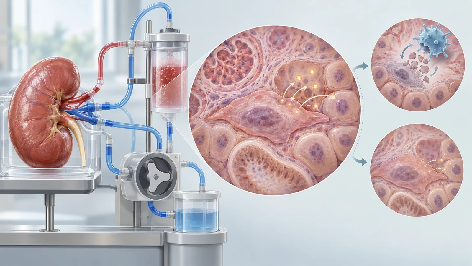
The proposed transplant application changes the setting of the experiment. Instead of giving a treatment throughout a recipient’s body, clinicians could potentially expose a donor kidney to a therapy while the organ is outside the body and connected to a machine-perfusion circuit before implantation.
The motivation comes from the stresses surrounding transplantation. Cold ischemia, ischemia and reperfusion injury, acute rejection and the procedure itself can promote oxidative stress, inflammation and vascular complications. The review connects these conditions with molecular changes that may accelerate graft aging and contribute to later decline.
Ex vivo perfusion creates a period in which the organ can be assessed or treated before it is placed in the recipient. The review presents senotherapy during this period as a possible way to improve graft quality. For a senolytic, the proposed benefit would depend on reaching harmful senescent cells while sparing viable tissue. For a senomorphic approach, it would depend on changing the inflammatory environment enough to help the kidney tolerate transplantation and recover.
The review also describes a separate vesicle-based experiment. Mesenchymal stem cell-derived extracellular vesicles were added to perfusion fluid, and the treated kidneys showed preservation of function in histological and genetic assessments. Vesicles are packages released by cells, so this approach is not interchangeable with a senolytic drug that selectively kills senescent cells. It is a distinct example of how perfusion fluid might be used to influence an organ before transplantation, not evidence that senolytic perfusion improved transplant outcomes.
What this could change
A successful senescence-directed perfusion treatment could eventually condition a donor kidney before implantation, giving a transplant recipient an organ better able to withstand procedural injury and retain function. That possibility depends on showing a real graft benefit while avoiding unacceptable damage to useful cells. In the April 2021 review, organ conditioning is a research proposal made visible by the biology of senescence, not an established transplant procedure.
Key Sources
- Primary source: Targeting Premature Renal Aging: from Molecular Mechanisms of Cellular Senescence to Senolytic Trials.
DOI or Journal Page · Review or Meta-Analysis · 2021-04-29