
Research Signal – Source date 2024-09-27
Chamomile’s familiar association with soothing effects took an unexpected turn in a laboratory experiment. In human skin fibroblasts previously exposed to the chemotherapy drug etoposide, chamomile extract sharply increased an inflammatory protein released into the surrounding liquid. Several genes associated with inflammatory signaling also became more active.
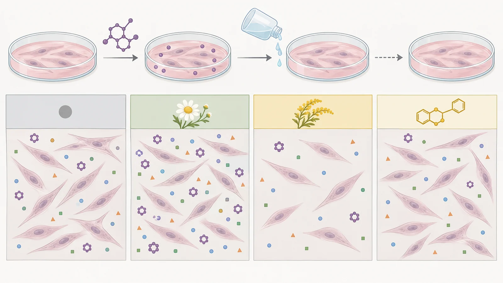
The original experimental study, published September 27, 2024, examined chamomile, goldenrod, green tea and reishi mushroom extracts, with quercetin as a comparison substance. Its most useful finding concerns two outcomes that can easily be confused in claims about herbal anti-aging treatments. A preparation can reduce the number of surviving cells without making the survivors less inflammatory. Conversely, it can alter inflammatory signaling without eliminating those cells.
That distinction helps explain why activity against senescent cells and increased inflammatory signals can appear within the same study. The experiments measured different aspects of the cells’ response, on different treatment schedules.
Cell survival and cell behavior are different targets
Cellular senescence is a state in which cells stop dividing after stresses such as DNA damage. They remain biologically active. Growth arrest can help prevent damaged cells from multiplying, while changes in what these cells release can affect surrounding tissue.
Those secretions include inflammatory proteins, growth factors and enzymes. Together, they are called the senescence-associated secretory phenotype, or SASP. Their effects depend on context; inflammatory signaling can contribute to healing as well as persistent tissue disruption.
Two approaches aim to change this situation. Senolysis eliminates senescent cells, ideally while sparing other cells. Senomorphic action changes their behavior, including potentially harmful secretions, without killing them. These are separate biological goals, so a result supporting one does not automatically support the other.
The study investigated both using primary human dermal fibroblasts, cells from the skin’s connective tissue. It compared extract effects in low-passage cells—cells that had undergone relatively few rounds of laboratory propagation—and cells exposed to etoposide to induce premature senescence.
Timing changed the question
The researchers used two schedules that matter for interpreting the results.
For toxicity testing, cells received etoposide for 48 hours. After the drug was removed, they spent another 12 days in culture before extract treatment began. This interval allowed the senescent state to develop before the researchers tested how the cells responded to different extract concentrations. They measured metabolic activity with PrestoBlue and used staining for senescence-associated beta-galactosidase, a commonly used marker of senescence.
In the chronic-treatment experiments, extract exposure began much earlier: the day after etoposide removal. Treatment continued while the cells developed their longer-term response to the damage. The team collected culture liquid and cellular RNA during this period to measure secreted IL-6, an inflammatory signaling protein, and the activity of selected genes.
The chronic experiments therefore examined how treatment affected the developing response to etoposide. They did not simply add extracts to a population that had already spent two weeks establishing senescence. This arm used two primary cell lines, with three independent experiments per line, giving the researchers a way to check whether the main patterns recurred across cell backgrounds.
Similar readings can have different explanations
Chamomile produced the clearest increase in inflammatory activity. IL-6 accumulated strongly in the culture liquid compared with etoposide-treated controls receiving no extract. Although the timing of the response differed between the two cell lines, the main increase appeared in both.
Gene-expression measurements supported a broader change. Chamomile increased RNA levels for IL-6, IL-8, IL-1α, IL-1β and CXCL-1, as well as MMP3, an enzyme involved in breaking down the material surrounding cells. These measurements concern the instructions cells produce for making proteins. They should be distinguished from the direct measurement of IL-6 protein in the culture liquid; the experiment did not measure every listed factor’s secreted protein level in the same way.
Goldenrod moved the IL-6 reading in the opposite direction, but microscopy supplied a crucial explanation. Treated cultures became much less densely populated. With fewer cells left to contribute IL-6, its concentration could fall even if each surviving cell continued releasing substantial amounts. The authors acknowledged that the goldenrod concentration may have been excessive for investigating senomorphic effects because most cells died. Its lower IL-6 reading therefore cannot establish an anti-inflammatory action independent of cell loss.
Quercetin also lowered IL-6 in both cell lines, which the authors interpreted as a senomorphic effect. Its gene-expression results were mixed, however. IL-1β expression decreased at all measured time points, while MMP3 expression increased. Reduced IL-6 did not mean that every part of the senescence-associated response had been suppressed.
Reishi’s contribution came from the toxicity and staining experiments. The mushroom extract was associated with a declining percentage of cells positive for the senescence-associated stain. That observation belongs to the evidence about viability and senescence markers; it does not supply an equivalent chronic IL-6 result.
Across the extracts, the authors interpreted differences in toxicity between senescent and low-passage cells, alongside staining observations, as evidence of senolytic activity. Those findings can coexist with chamomile’s inflammatory response because susceptibility to an extract and inflammatory output are different measurements. The separate experiments do not trace the fate and secretion of each individual cell.
Antioxidant strength did not predict the response
The team also tested a common explanation for the appeal of plant extracts: their antioxidant properties. A chemical assay measured radical-scavenging activity, and a separate assay assessed polyphenol content.
Green tea had the strongest antioxidant activity among the extracts, approaching that of the antioxidant control, Tempo. Yet it did little to reduce the senescence-associated inflammatory response measured through IL-6. Its IL-6 readings were close to those of cultures receiving no extract in both cell lines. Tempo likewise did not produce the desired suppression.
Polyphenol content also failed to show a straightforward relationship with senolytic or senomorphic effects. Green tea was rich in these compounds, but it was toxic to both low-passage and etoposide-treated cells at the tested concentrations.
A chemical radical-scavenging assay measures one property of a preparation. The cells’ response also involves survival and regulation of inflammatory activity. In this experiment, antioxidant strength alone could not predict which of those outcomes would follow.
How far the experiment can travel
Several features limit what can be inferred about selective cell removal. PrestoBlue measures metabolic activity, which is used as an indicator of viability. A lower reading can reflect fewer viable cells, altered activity in the remaining cells, or a combination. Microscopy and senescence staining add useful information, but differential viability in this model does not establish a safe therapeutic window across human tissues.
For the chronic experiments, the authors explicitly acknowledged that they did not measure exact cell numbers. The images showed changes in density and shape, but they could not provide a precise calculation of IL-6 release per surviving cell. This is especially consequential for goldenrod, where extensive cell loss accompanied the lower protein concentration.
The model also captures a particular form of stress. Etoposide damages cells and induces premature senescence; that process does not reproduce every feature of cells aging through repeated division or of aging tissue inside a person. Directly bathing fibroblasts in extracts also differs from the exposure produced by digestion, metabolism or application to skin. The study supplies no basis for equating its culture concentrations with those reached through ordinary product use.
These boundaries are essential for interpreting chamomile. Its mechanism remained unresolved, and the authors called for confirmation in living organisms. The findings do not establish that drinking chamomile tea causes a clinical cytokine storm or a harmful interaction with chemotherapy. Nor do the extract results demonstrate longer life, improved health or reversal of aging in people.
What this could change
If compounds from these preparations can eventually alter senescent cells selectively at exposures achievable in living tissue, they could contribute to treatments that preserve tissue function by reducing persistent inflammatory signaling or removing cells that sustain it. The distinction between those approaches would matter clinically: changing a cell’s secretions could offer a way to reduce harmful effects while retaining the cell, whereas selective removal could address a population whose continued presence causes damage.
That possibility depends on connecting the cellular effects to better tissue function while preserving useful stress responses and repair. This study contributes an early experimental distinction needed for that goal. It shows why a promising preparation must be understood through both the cells that remain and what those cells continue to release.
Key Sources
- Primary source: Exploring Senolytic and Senomorphic Properties of Medicinal Plants for Anti-Aging Therapies.
DOI or Journal Page · Peer-Reviewed Paper · 2024-09-27
Keep exploring