
Evidence date · 2022-09-26 · Research note 1 of 4
1 2 3 4
See current Track status →
Memory and thinking can deteriorate because another illness disrupts the brain. Sometimes, identifying that cause opens an opportunity to treat it. Normal-pressure hydrocephalus illustrates the stakes. It can combine difficulties with walking, cognition, and bladder control, and surgical placement of a shunt to divert fluid can improve walking and cognition in some patients. Recognizing a treatable cause matters, even though the term “secondary” does not mean every cognitive disorder can be reversed.
A review published on September 26, 2022, examined what transcranial magnetic stimulation, or TMS—magnetic pulses used to stimulate the brain—could contribute to these problems. It brought together a broader literature on measuring brain physiology and following responses to treatment, alongside a few experiments that tried to improve cognitive performance directly.
The authors searched PubMed from its inception through July 14, 2022. Two authors independently screened titles and abstracts, with a third resolving disagreements, and they checked references for additional studies. Their scope included conditions such as multiple sclerosis, thyroid disease, and sleep disorders; having one of these conditions does not itself mean someone has dementia. They excluded vascular cognitive impairment and cognitive disorders attributed to psychiatric diseases or specified neurosurgical conditions.
From a magnetic pulse to a muscle response
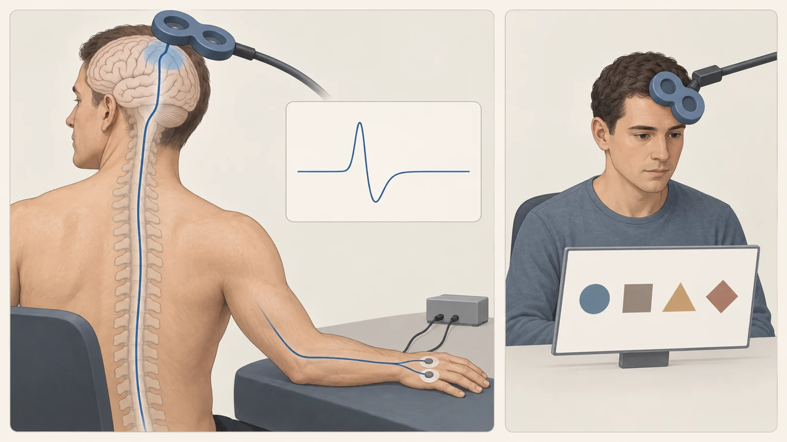
Much of the work begins over the motor cortex, a brain region involved in movement. A coil delivers a magnetic pulse over this area, triggering activity that travels through motor pathways toward a muscle on the opposite side of the body. Researchers record the resulting electrical response, called a motor evoked potential. A hand muscle can thus provide a measurable endpoint for an experiment that starts at the brain.
Changing the pulse strength helps establish the resting motor threshold, the minimum stimulation intensity needed to elicit a response under resting conditions. Researchers can also estimate how long activity takes to travel along the pathway from the brain through the spinal cord, accounting for the peripheral part of the journey. These measurements reveal different aspects of responsiveness and conduction. Their values reflect contributions from several components, including cortical circuits, spinal motor neurons, peripheral nerves, and muscle.
Other tests investigate inhibition, the processes that restrain neural activity. Short-latency afferent inhibition, known as SAI, probes circuitry involved in the interaction between sensory input and motor activity. It is sensitive to acetylcholine, a chemical messenger, but other neurotransmitters, including GABA, can influence it too. That makes it a window onto interacting circuits rather than a direct assay of one brain chemical.
Why examine movement pathways when the concern is memory? The review explains that illnesses affecting cognition can also involve motor areas, either through the disease process itself or through changes in how the brain’s networks operate.
Different illnesses alter the balance of activity
The reviewed conditions did not all push the motor cortex in the same direction. Studies of normal-pressure hydrocephalus found increased excitability and reduced inhibition associated with GABA signaling. Obstructive sleep apnea, by contrast, tended to show greater motor cortex inhibition overall. Chronic insomnia and sleep deprivation were associated with less inhibition. Even among conditions involving sleep, “more brain activity” could not describe one consistent therapeutic goal.
Increased excitability also had more than one possible explanation. The authors proposed that it might sometimes be a compensatory response to injury, helping preserve circuit function. Alternatively, it could reflect preferential damage to inhibitory neurons, leaving activity less restrained. The same broad observation could therefore arise from different biological processes.
That distinction matters when considering stimulation intended to change brain activity. Reducing excitability might be worth testing when inhibitory control is impaired; a compensatory increase would call for a different interpretation. The authors discussed both lower- and higher-frequency stimulation approaches matched to different physiological patterns. Those proposals remained research hypotheses, with no validated method for choosing a cognitive treatment from an individual’s TMS profile.
Recovery can follow more than one timetable
Treatment of an underlying illness offered another way to examine these circuits. A 2008 study summarized in the review compared 10 people with overt hypothyroidism with 10 healthy participants of similar ages. Before hormone replacement, the patients showed several changes, including a higher motor response threshold and altered inhibition. After three months of treatment, most measured parameters had returned to normal. One measure of local cortical inhibition, called short-interval intracortical inhibition, took six months to normalize. TMS was tracking the physiological response to hormone replacement in this experiment. The different timings showed how components of the same system could recover at different rates.
The experiments that tried to improve memory
Repetitive TMS, or rTMS, delivers sequences of pulses to influence circuit activity. A few studies in the review used it to test changes in cognitive performance.
In a 2017 multiple-sclerosis study, the review’s table lists 17 patients and 11 healthy controls. Researchers stimulated the right dorsolateral prefrontal cortex, a frontal brain region, at 10 pulses per second. The review reports improved working-memory task accuracy after real stimulation but not after sham stimulation, the comparison intended to imitate treatment. Working memory is the ability to hold information briefly and use it during a task.
The researchers also found increased task-related functional connectivity between the stimulated frontal region and the right caudate nucleus and parts of the cingulate cortex. This describes changes in how activity in those regions was related during the task. The authors interpreted the findings as suggesting a change in network efficiency.
A separate 2013 study concerned healthy volunteers undergoing sleep deprivation. It included 27 sleep-deprived participants receiving real or sham stimulation and 21 participants who were not sleep deprived. Four stimulation sessions were delivered over a two-day period of sleep deprivation. The actively stimulated, sleep-deprived group did not show slowing or lapses and performed similarly to participants who had not been deprived of sleep. The review also reports improved working-memory performance 18 hours after the final session.
These studies tested cognitive performance directly, going beyond a change in a physiological measurement. Their interpretation nevertheless depends on who was tested and what improved. Sleep-deprived healthy volunteers are a different population from people with chronic cognitive disorders, and the 18-hour observation belongs to that particular sleep-deprivation experiment. These findings do not establish lasting gains in everyday functioning, prevention of dementia, or a treatment effect that applies across secondary cognitive disorders.
What a useful clinical test would have to resolve
As of the evidence assembled in July 2022, the authors could identify no distinctive TMS pattern ready to predict whether cognitive decline would occur or progress in a particular condition. A central difficulty was separating the effects of an illness on the nervous system from the changes specifically associated with cognitive impairment.
Multiple sclerosis illustrated this problem. Some studies linked TMS findings to cognition, including a study in which reduced SAI was associated with cognitive dysfunction. Yet another study of 28 people with primary progressive multiple sclerosis found that evoked potentials, including motor responses, were moderately related to clinical and functional status without an association with cognitive impairment. A measure can be informative about the course of a neurological disease while having a different, less certain relationship to memory and thinking.
Patient selection therefore matters. Many studies included in the review did not specifically select participants according to the presence and measured extent of cognitive impairment. Comparisons within the same disease, involving people with and without cognitive difficulties, would help determine what an abnormal response means. Following participants over time would then be necessary to test whether it predicts later decline.
The studies were also generally small and covered heterogeneous conditions, which the authors identified as obstacles to combining results. Even repeatedly observed abnormalities could be affected by publication bias, with some kinds of results more likely to reach the literature than others. Repetition across small reports alone could not settle clinical usefulness.
Comparability between centers was another practical barrier. The review called for standardized TMS procedures and more consistent interpretation of motor responses. If measurements are to support decisions across clinics, researchers need confidence that differences between patients reflect biology rather than differences in collection or analysis. For stimulation treatments, that effort also needs to connect reproducible circuit findings with meaningful cognitive outcomes.
What this could change
If researchers can validate disease-specific circuit targets and show that stimulation produces lasting, meaningful gains in cognition, people whose memory and thinking are affected by another illness could gain an additional treatment alongside care for the underlying condition. The benefit would be help with cognitive difficulties that affect everyday life.
Reliable brain-response measurements could support that treatment by helping researchers identify relevant circuits and follow their response over time.
Key Sources
- Primary source: A comprehensive review of transcranial magnetic stimulation in secondary dementia.
DOI or Journal Page · Peer-Reviewed Paper · 2022-09-26