
Research Signal – Source date 2025-08-27
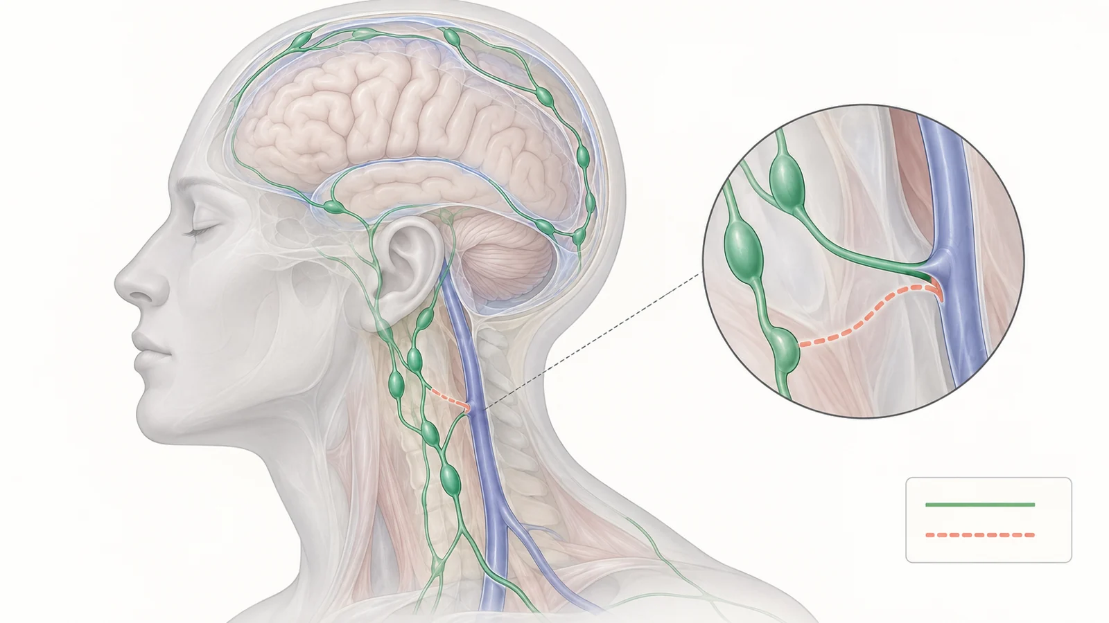
An operation aimed at Alzheimer’s disease might seem certain to involve the brain. An emerging surgical approach instead targets tiny lymphatic vessels deep in the neck, creating connections to nearby veins. The intention is to influence an outlet through which fluid and dissolved material drain from the head.
A systematic review published August 27, 2025, examines this approach. Its authors searched PubMed and the Cochrane Library, supplemented by Google Scholar, in July 2025, restricting their search to publications from 2015 onward. They identified nine publications addressing the surgical approach—a small, varied literature that brings together anatomy, experimental reasoning, and early patient observations.
Why the neck enters the picture
The proposed connection starts with two fluids. Cerebrospinal fluid surrounds the brain and spinal cord; interstitial fluid occupies spaces between brain cells. Exchanges between these fluids help move dissolved substances, including products of cellular activity. The glymphatic model describes exchange involving spaces around blood vessels and specialized support cells called astrocytes.
Lymphatic vessels in the meninges, the coverings around the brain, form another part of the drainage network. They provide routes toward deep cervical lymph nodes in the neck. Fluid exchange within brain tissue and lymphatic drainage outside it are connected processes, but they occur in different anatomical compartments. The operation targets the cervical portion of this distributed system.
The review also describes intramural periarterial drainage, or IPAD, an alternative model in which substances leave along structures within capillary and arterial walls. These descriptions should not be compressed into a single settled circuit. Their relevance to surgery is the broader possibility that events at a downstream drainage route could influence clearance farther upstream.
Why try to alter that drainage? The authors discuss declining clearance with aging and experiments in mice in which researchers disrupted lymphatic pathways genetically, chemically, or by tying off cervical vessels. Such interventions impaired removal of disease-associated proteins, including amyloid-beta, phosphorylated tau, and alpha-synuclein. Some experiments also found cognitive deficits.
Those experiments establish what happened when drainage was disrupted in animals. They do not establish the effect of adding a surgical connection in a person with Alzheimer’s disease. The authors’ description of impaired clearance as “cerebral lymphedema” is a proposed therapeutic framing, not a diagnosis demonstrating that every patient has the same drainage problem. Alzheimer’s disease cannot be reduced to a blocked outlet.
What surgeons connect
An anastomosis is a surgically created connection. In lymphatico-venous anastomosis, usually shortened to LVA, a lymphatic vessel is joined to a nearby vein or venule. The aim is to give lymph another route into the venous circulation. The review describes supermicrosurgery capable of joining vessels less than 0.8 millimeters across.
LVA already has an established use in peripheral lymphedema, where impaired lymph transport causes swelling outside the brain. Applying the technique to deep cervical drainage brings it into a different physiological setting. The proposed neck operation does not directly remove protein deposits from brain tissue; it changes a connection farther along the drainage network.
The reviewed procedures also differ from one another. One report used an “Octopus technique” joining several small lymphatic vessels to one vein, together with lymph-node-to-vein connections. Another described a modified procedure involving a flap containing deep cervical lymphatic vessels and nodes, connected to the external or internal jugular vein. A further report described an end-to-end vessel connection.
A lymphatic-vessel-to-vein bypass and a lymph-node-to-vein connection therefore should not be treated as interchangeable descriptions of one standardized operation. Technique variation matters when interpreting the patient reports: different reconstructions may have different flow characteristics, even when grouped under the same therapeutic idea.
What the early patient reports measured
The review’s nine publications are not nine independent clinical trials. Its catalog includes case reports, a prospective single-arm cohort, and a narrative review. The authors describe substantial differences in surgical methods, outcome measures, and follow-up periods, so the reported changes cannot be read as a pooled estimate of benefit.
Across the clinical reports, early postoperative improvements commonly appeared within the first month. Changes on the Mini-Mental State Examination, or MMSE, ranged from 2 to 5 points; some reports also described gains of up to 2 points on the Montreal Cognitive Assessment, or MoCA. These are cognitive assessment scores. A rise records better performance on the assessment at that visit, without by itself establishing restored independence or a lasting change in disease progression.
One prospective cohort discussed in the review involved 26 patients receiving a modified procedure. At one month, the investigators reported a statistically significant 2-point MMSE increase. The MoCA findings were less clear, with improvement reported in a minority of patients without statistical significance. Different tests thus did not provide an identical picture of change.
The central attribution problem is the missing comparison in a single-arm study. Measuring patients before and after an operation shows a sequence of events, but cannot isolate what surgery contributed. Repeat testing, fluctuations in performance, and other aspects of treatment or care remain possible influences that such a design cannot separate. Statistical significance within a treated group does not answer what would have happened to comparable patients without the operation.
Everyday function and behavior were assessed separately. Some reports described changes on measures of daily activities, depression, or neuropsychiatric symptoms. These observations address different questions from cognitive testing, including how patients function and feel. They should retain their separate identities rather than being folded into a general claim of recovery.
Biological measurements had still other time scales. In a subset of 18 patients from the cohort, cerebrospinal-fluid biomarkers showed a downward trend 5–7 days after surgery. The review also describes imaging reports of increased glucose metabolism and changes interpreted as reduced disease-associated protein burden. An early biomarker change, an imaging finding, and a better cognitive score are different observations; the synthesis does not establish that one caused another or that they predict durable benefit.
Although Parkinson’s disease appears in the review’s broader discussion, the detailed patient outcomes considered here concern Alzheimer’s disease. They do not provide comparable clinical evidence for Parkinson’s treatment.
A connection needs the right pressure
Joining two vessels does not guarantee useful flow. The proposed bypass depends on a favorable pressure relationship that allows lymph to enter the receiving vein. For a neck connection to improve brain drainage, the relevant upstream compartments must also communicate in a way that supports that movement.
The review gives an estimated cervical venous pressure range of 0–6 millimeters of mercury, but emphasizes that precise measurements of the relevant human cerebral lymphatic pressure gradient are lacking. Mouse experiments support pressure-driven movement between the space around the brain and cervical lymphatics. Whether the necessary relationship exists in the human system remains unresolved. A technically successful junction therefore does not, by itself, demonstrate improved clearance throughout the pathway.
Pressure also introduces a distinct safety question. If venous pressure favors movement in the opposite direction, blood could reflux into the lymphatic connection. The authors discuss possible consequences of backflow toward the central nervous system and uncertainty about the presence and function of valves in human meningeal lymphatics. These are physiological concerns, not evidence that a particular rate of such complications has been measured in these patients.
Techniques intended to limit reflux exist in peripheral lymphatic surgery, and the review discusses them. Their performance in that setting does not demonstrate protection of the cerebral drainage network. Evaluating the cervical approach requires evidence about flow direction and safety in the anatomy actually being treated.
What this could change
The consequential clinical question is whether adding surgery to pharmacological treatment gives patients a better outcome than pharmacological treatment alone. That comparison asks more than whether someone scores higher after an operation. It tests the extra contribution of surgery against care that both groups receive.
The review’s registry table records a planned multicenter randomized study with that comparison: 92 participants, divided into two groups of 46, with cognitive function and quality of life among the outcomes. It also lists an exploratory study with 10 participants in a single group. These entries describe research plans preserved in the review, not completed results. The designs serve different purposes: a single-group study can characterize postoperative changes, while a randomized comparison is better suited to estimating the added effect of the intervention.
For patients and families, the meaningful result would be a sustained difference in cognition or everyday life that justifies the burdens and risks of an operation. Follow-up would need to show how long any advantage lasts, while safety assessment captures consequences that a brief cognitive test cannot. Measurements of drainage could help explain why an effect occurs and which patients might experience it.
If comparative studies established that combination of benefit and acceptable safety, deep cervical drainage could become a treatment target beyond the brain itself. The important advance would be evidence that changing this accessible part of the lymphatic network makes a lasting difference to people living with Alzheimer’s disease.
Key Sources
- Primary source: Brain lymphatic drainage pathways, deep cervical lymphatic surgery, and current insights: A systematic review.
DOI or Journal Page · Review or Meta-Analysis · 2025-08-27
Keep exploring