
Research Signal – Source date 2026-07-01
A two-drug treatment was linked to several changes in the kidneys of aged mice: fewer signs of cell senescence, less scarring, and less fat accumulation in kidney tubules. The findings also extended to gene activity across kidney cell types and to computational estimates of how those cells communicate.
But tissue changes do not answer the most practical question: whether the kidneys worked better. The researchers did not prospectively collect blood or urine measures of kidney function in this long-term mouse study. The results describe changes in kidney tissue and molecular patterns, not a demonstrated improvement in filtration or protection from kidney disease.
The study, published July 1, 2026, tested dasatinib plus quercetin, often abbreviated D + Q, in naturally aged male mice. It offers a detailed look at how treatment-associated changes appeared across the kidney, while leaving their functional importance open.
Eight months of treatment changed several tissue features
The researchers compared three groups: young mice, old mice given a vehicle solution, and old mice given D + Q. The mice were randomly assigned to groups, and investigators were blinded to group assignments during baseline characterization, treatment, and outcome assessments. Treatment began when the mice were 12 months old and continued until they were 20 months old. The study examined their kidneys at the end of that period.
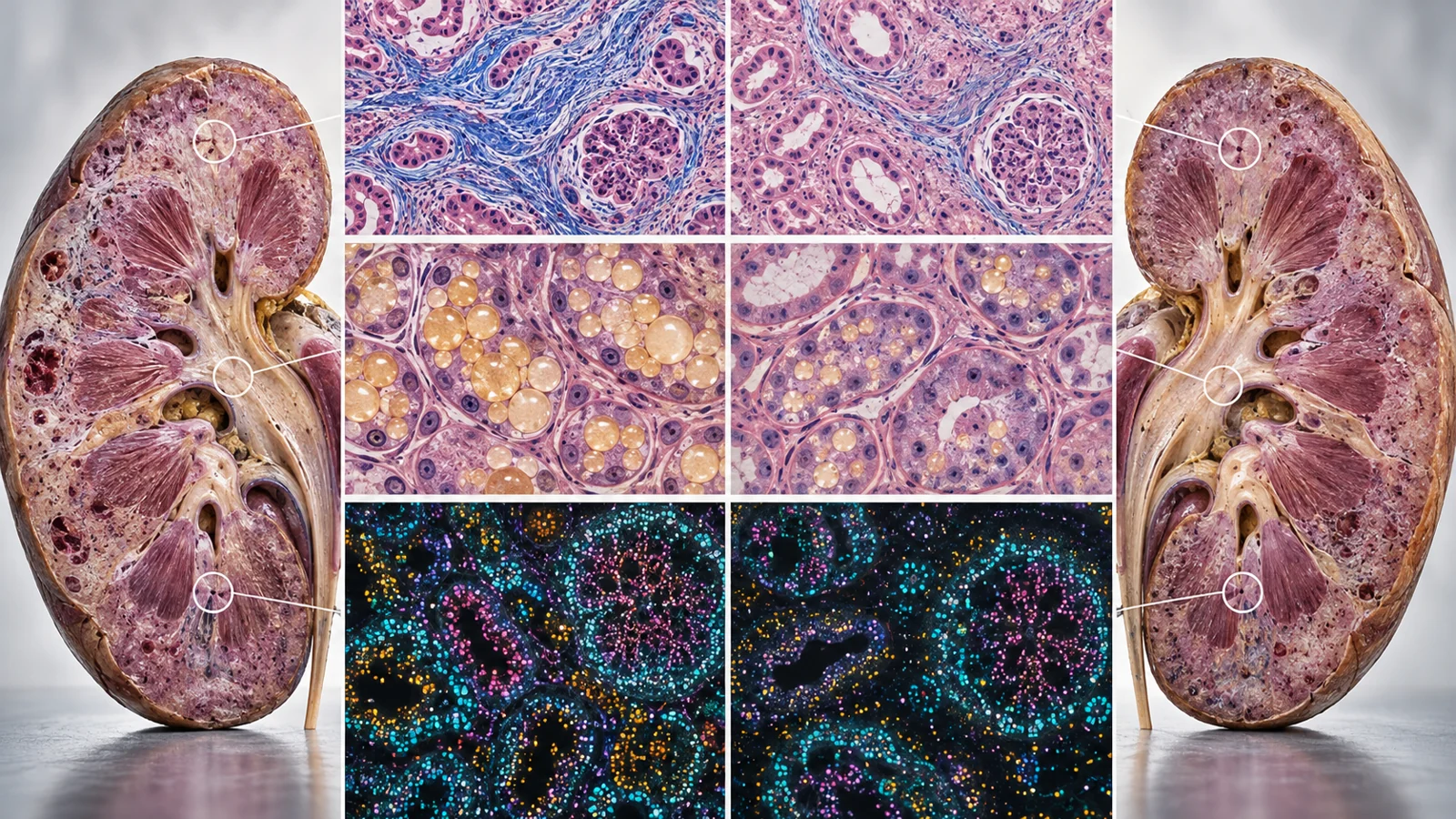
Senescent cells are cells that have stopped dividing but remain active in ways that can affect nearby tissue. In aged kidneys, the researchers found more p16 and p21, proteins associated with senescence, as well as more senescence-associated beta-galactosidase staining. In treated old mice, measurements of these markers were lower than in vehicle-treated old mice. Staining for Klotho, a protein associated with kidney aging, was also partly restored.
The tissue findings went beyond those markers. Aged kidneys showed more signs of structural injury, including fibrosis: the buildup of scar-like material in tissue. D + Q-treated kidneys had less staining for alpha-smooth muscle actin and Collagen I, and less Masson’s trichrome-positive area, measures the study used to assess fibrotic remodeling. Staining linked to inflammation also fell. The treated kidneys showed partial restoration of WT-1 staining, which the researchers used to assess cells in the glomerular compartment, the kidney’s filtering structures.
These are measurements of tissue appearance and molecular markers. They show that the differences between treated and vehicle-treated aged mice were not confined to senescence markers. They do not by themselves establish that removing senescent cells caused each other change: D + Q can affect multiple pathways, and the study’s measurements do not assign a complete cause-and-effect order among them.
The lipid findings point to altered fuel handling
Kidney cells use fatty acids as an energy source. The study focused on PPARα, a regulator of genes involved in fatty-acid use. In aged kidneys, the researchers found lower levels of PPARα and two related proteins, CPT1A and ACOX1. After D + Q treatment, levels of all three were higher than in vehicle-treated aged kidneys.
The researchers also saw fewer lipid droplets and less vacuolization in tubular cells, which line much of the kidney’s filtering and reabsorbing structures. Staining for 4-hydroxynonenal, a product associated with lipid peroxidation, was lower as well. Proteomic analysis found enrichment of lipid-metabolism processes after treatment, consistent with the staining results.
Together, these measurements suggest that aged kidneys treated with D + Q had a different pattern of lipid handling and less evidence of lipid accumulation and damage. They do not directly measure how much fatty acid the kidneys used or how quickly it was metabolized. Nor do they show whether changes in lipid handling came before, after, or independently of the changes in senescence, inflammation, or scarring.
Gene patterns shifted differently across kidney cells
A kidney contains many cell types, and an overall tissue measurement can obscure how each responds. To look more closely, the researchers used single-cell RNA sequencing, which measures gene activity in individual cells. They identified major kidney populations including proximal tubule, endothelial, interstitial, fibroblast, and podocyte cells.
The analysis found aging-related gene-expression patterns across several cell types. A transcriptional aging clock, built from gene-expression patterns, predicted higher cellular ages in old kidneys than in young ones. In treated old mice, its predicted ages shifted lower in several cell types, including proximal tubule, endothelial, interstitial, fibroblast, and podocyte cells.
Those “ages” are model-derived predictions, not direct measurements of how old a cell is or proof that a cell has been biologically rejuvenated. The clock inferred an age-like pattern from gene expression. Its value here is comparative: it showed that treatment-associated shifts were visible across multiple cell populations rather than only in a whole-kidney average.
The pathways associated with those shifts were not identical across cell types. In proximal tubule cells, treatment-related gene changes were enriched for fatty-acid metabolism. In endothelial cells, the analysis highlighted pathways related to response to transforming growth factor beta, a signaling molecule involved in tissue remodeling. In interstitial cells, it identified changes linked to tight-junction integrity, Rap1 signaling, and peroxisome function. The cell-specific results add detail to the tissue findings: the kidney did not respond as one uniform compartment.
Fewer predicted interactions do not show messages passing
Cells influence one another through signals. The researchers used CellChat, a computational tool that infers possible cell-to-cell signaling from the expression of ligand and receptor genes. In aged kidneys, the analysis predicted more interactions and stronger overall communication between cell populations than in young kidneys. D + Q-treated aged kidneys showed fewer predicted interactions and lower overall interaction strength than vehicle-treated aged kidneys.
The analysis also found changes in predicted APP, visfatin, and BMP signaling networks, which the paper discusses in relation to inflammation. These networks are inferred from gene-expression data and a database of ligand-receptor pairs. They suggest that the pattern of potential communication changed; they do not directly observe molecules being sent between cells or establish which signal caused a tissue outcome.
The key functional boundary remains straightforward: the long-term cohort did not include prospectively collected blood and urine endpoints to test kidney function. The authors therefore could not determine from this experiment whether the tissue and molecular differences translated into improved filtration or other functional benefit. A single study in male mice also cannot establish benefit or long-term safety for people with aging kidneys.
What this could mean for kidney protection
If later studies show that the changes in scarring, lipid buildup and inflammation preserve kidney function, and that treatment is safe in people, senolytic therapy could one day help older adults maintain kidney function as they age. This study does not establish that benefit: its aged-mouse cohort had no prospective kidney-function measurements, and it cannot establish long-term safety in people.
Key Sources
- Primary source: Multi-omics profiling reveals systemic rejuvenation of the aged kidney through senolytic therapy.
DOI or Journal Page · Peer-Reviewed Paper · 2026-07-01
Keep exploring