
Research Signal – Source date 2021-01-01
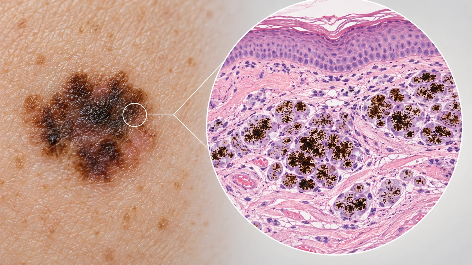
A new dark lump or patch in someone being treated for metastatic melanoma can look like another deposit of cancer. Yet a biopsy of a melanoma-like lesion may show a different picture: pigment-filled cleanup cells, with no viable melanocytes in the sampled tissue. That pattern is called tumoral melanosis.
The finding can be an important clue about what happened in the tissue that was sampled. It cannot, by itself, tell clinicians whether melanoma remains elsewhere in the body. A 2021 report of 10 patients treated with immunotherapy illustrates both why the pattern drew attention and why it still needs careful interpretation.
A dark lesion can contain pigment without viable melanoma cells
Melanocytes are the cells that make pigment. When melanoma cells break down, pigment can remain in the tissue and be taken up by melanophages, immune cells that contain and help clear pigment. Under the microscope, tumoral melanosis appears as a nodular collection of these melanophages without viable melanocytes in the sampled area.
To the eye, however, the lesion can resemble metastatic melanoma. The mismatch between appearance and biopsy is the puzzle: a suspicious-looking spot may reflect a local tissue change after treatment rather than a deposit of living melanoma in that particular sample. The biopsy finding still calls for evaluation to rule out melanoma in the lesion or metastatic disease elsewhere.
The report focused on patients with metastatic melanoma who developed tumoral melanosis after immunotherapy. The treatments included drugs that act on immune checkpoint proteins, which normally help regulate immune activity. The authors described the pattern as a possible sign of treatment-related tumor regression, while noting that its biological basis and prognostic meaning were not yet clear.
The finding appeared months into treatment, with exceptions
In this retrospective series from one tertiary care institution, the 10 patients developed tumoral melanosis between 2 and 20 months after starting immunotherapy. The mean time was 10 months. At follow-up, eight patients were classified as having a complete or partial response to treatment. That combination—melanoma-like lesions appearing during treatment and many patients also being classified as responders—helped make the pattern clinically interesting.
The outcomes were not uniform. One patient’s melanoma progressed in internal organs and the skin despite tumoral melanosis. Another developed new cancer deposits in the abdominal lining and skin despite the finding. One patient had not yet had repeat imaging, so the status of disease elsewhere remained unassessed at that point.
The report also records three patients alive with no active disease at follow-up and five alive with disease. Among those alive with disease, two had nearly complete responses before cancer returned in internal organs after immunotherapy stopped. This range of outcomes shows why a biopsy finding in one lesion cannot stand in for assessment of disease throughout the body.
What a 10-patient report can establish
The study was a retrospective case series, not a prospective test of whether tumoral melanosis predicts response. It records a pattern in 10 patients and describes their timing and clinical outcomes. Because the series is small and comes from one institution, it cannot establish how often the finding occurs among people receiving immunotherapy, how reliably it corresponds to regression, or whether it predicts a lasting response.
Nor does the absence of viable melanocytes in one sampled lesion rule out melanoma in that tissue beyond the sample or metastatic disease at another site. The progression described in the report makes that limit concrete. The authors called for larger prospective studies and longer follow-up to clarify the finding’s frequency and its relationship to cancer outcomes.
What tumoral melanosis could add to melanoma follow-up
If larger prospective studies show that this biopsy pattern reliably relates to disease status elsewhere, tumoral melanosis could give clinicians an additional clue when interpreting suspicious lesions during melanoma follow-up. Used alongside broader assessments of disease, it might help distinguish a pigment-rich, regressed tissue site from a lesion containing viable melanoma. For now, this small series raises that possibility; it does not establish the finding as a dependable measure of treatment success.
Key Sources
- Primary source: Tumoral Melanosis in the Setting of Targeted Immunotherapy for Metastatic Melanoma-A Single Institutional Experience and Literature Review.
DOI or Journal Page · Review or Meta-Analysis · 2021-01-01
Keep exploring