
Research Signal – Source date 2022-10-05
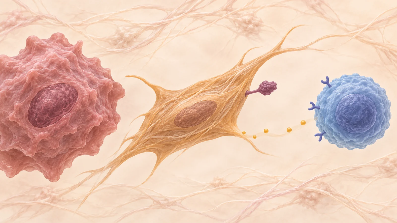
Checkpoint drugs are designed to block inhibitory signals that can keep immune cells from attacking cancer. But those signals may involve more than the cancer cells themselves. In a 2022 review, researchers describe how cancer-associated fibroblasts—support cells within tumors—can display checkpoint ligands or influence which checkpoint molecules appear on nearby immune and cancer cells.
A checkpoint molecule is part of the immune system’s braking machinery. Some checkpoint drugs block these inhibitory interactions, helping restore immune-cell activity. The review’s central point is that the cells shaping those interactions can vary: a fibroblast may carry a ligand itself, or release substances that affect other cells. The examples come from different cancers and research settings, so they do not add up to one universal pattern or establish a new patient treatment.
How ordinary fibroblasts become part of a tumor
Fibroblasts are connective-tissue cells that help maintain tissue and support repair. In a tumor, local conditions and signals from neighboring cells can activate some fibroblasts. The review describes factors such as inflammation, oxidative stress, low oxygen, and growth signals as contributors to this change. Activated fibroblasts are called cancer-associated fibroblasts, or CAFs.
CAFs are not a single uniform cell type. They can differ in origin, features, and behavior, even within the broad category. The review discusses markers used to identify them, including FAP and α-smooth muscle actin, but no single marker defines every CAF in every tumor. This variation matters because the immune effects attributed to fibroblasts depend on which cells are present and what they are doing.
Within a tumor’s surrounding tissue, CAFs can provide physical support and release signaling substances. Some of those substances can affect immune cells directly; others can prompt cancer or immune cells to change their own checkpoint expression. The location of a checkpoint signal therefore matters as much as its name.
A checkpoint ligand can sit on the support cell
In head and neck squamous cell carcinoma samples, the review describes CAFs expressing the checkpoint ligands PD-L1 and PD-L2, unlike normal fibroblasts in the comparison. The CAFs and their secretions also suppressed T-cell proliferation more strongly than normal fibroblasts in the reported study. This example places the ligands on the surrounding support cells, rather than only on cancer cells.
A different pattern appears in the review’s ovarian cancer example. B7-H3 was found on both tumor and stromal cells, with higher levels on stromal cells than on cancer cells. The comparison shows why a tissue sample’s overall checkpoint level may not tell us which cell type is carrying the signal. The review also notes that B7-H3 findings differed across tumor settings, underscoring that location cannot be assumed from one cancer to another.
These are observations from particular tumor samples and studies summarized by the review. They do not show that measuring a fibroblast ligand can already guide treatment for an individual person. They do show why researchers distinguish the checkpoint on a cancer cell from the same or another checkpoint on nearby stromal cells.
Secreted factors can change the brakes on other cells
Fibroblasts may also affect checkpoint patterns without displaying the relevant molecule themselves. In the reviewed examples, CAF-derived substances were associated with changes in checkpoint expression on T cells or cancer cells. The effects differed by tumor type and by the cell being measured.
For example, melanoma CAFs produced arginase, an enzyme that can alter the local chemical environment. In the studies summarized, exposure to these CAF-related factors was linked to increased TIGIT and BTLA on CD8 T cells, a type of immune cell that can kill abnormal cells. By contrast, the review describes pancreatic CAF-derived secretions as decreasing TIGIT on CD8 T cells. The direction of change was not the same in these two settings.
Other examples concern cancer cells. In studies of melanoma and colorectal cancer models, CAF-derived CXCL5 was linked to increased PD-L1 on tumor cells through a cellular signaling pathway. In a separate set of mouse tumor findings, CAF-associated NOX4 was linked to increased CTLA-4 on CD8 T cells; inhibiting NOX4 restored response to immunotherapy in those models. These results illustrate possible routes from fibroblast activity to checkpoint expression, but they come from different experimental contexts and should not be read as a single tested treatment strategy.
The review also describes pancreatic CAFs as promoting several checkpoint molecules on proliferating T cells, including PD-1, CTLA-4, TIM-3, and LAG-3, alongside reduced immune function in the reported studies. Such lists can sound like a standard signature, but the melanoma and pancreatic TIGIT examples point in different directions. A fibroblast effect observed in one tissue does not establish what will happen in another.
Where a signal appears changes its meaning
Checkpoint expression can reflect a complicated exchange among fibroblasts, cancer cells, and immune cells. The review describes CAFs as capable of directly bearing inhibitory ligands and indirectly influencing checkpoint expression through secreted factors. It also notes that the pattern may depend on tumor type, fibroblast subset, and the cell on which a marker is measured.
The evidence spans human tumor tissue, cell-based experiments, and animal models. These approaches can reveal where a molecule appears or how cells respond under studied conditions. They do not, by themselves, establish that a CAF-directed add-on improves patient outcomes or that a checkpoint pattern is a validated test for choosing treatment. The review organizes prior findings; it does not report a new clinical intervention testing that proposition.
The distinction is relevant to proposals discussed by the authors, such as removing CAFs or changing their activity toward a less tumor-supportive state. Fibroblasts have varied roles, and the reviewed examples do not establish that broadly targeting them would help. Any proposed approach would need to be studied in the relevant cancer, fibroblast population, and immune-cell context. Existing checkpoint drugs already target some immune brakes; the open question here is whether knowing where those brakes arise could add useful, validated information.
What this could make possible
One day, a tumor biopsy might be interpreted for immune-braking signals in both cancer cells and the fibroblasts around them. If researchers can validate which cell-specific patterns matter and show how they inform treatment decisions, that fuller picture could give clinicians a more dependable basis for studying—and eventually choosing among—checkpoint approaches. The review provides an early evidence bridge: fibroblasts can carry or influence different checkpoint signals in different tumors. Better measurement, prediction, and clinical testing would still be needed before that capability could guide care.
A nearer milestone would be independent studies in people showing that fibroblast-specific checkpoint patterns predict response to existing checkpoint drugs beyond what tumor-cell measurements alone can tell us. The review’s examples make that question worth investigating, while leaving its answer open.
Key Sources
- Primary source: Immune checkpoint inhibitors as mediators for immunosuppression by cancer-associated fibroblasts: A comprehensive review.
DOI or Journal Page · Review or Meta-Analysis · 2022-10-05
Keep exploring