
Research Signal – Source date 2022-09-26
A porous structure can give living cells somewhere to settle without providing everything they need to repair damaged tissue. Its surface may discourage attachment. It may release an ingredient too quickly, or remain in place longer than intended. In tissue engineering, this supporting structure is called a scaffold. Making one that accommodates cells is an important step; making one that helps tissue form requires several functions to work together.
A review published on September 26, 2022, brings those functions into focus through prior research on materials for tissue engineering. Its examples range from laboratory measurements and cell cultures to implants tested in animals. Read together, they explain what researchers can change in a scaffold—and why a favorable result in one test answers only part of the repair question.
Where the cells enter the process
The review describes two approaches that differ in when and where cells meet their support. In the first, called ex vivo tissue engineering, researchers seed cells onto a scaffold outside the body. They maintain it in a controlled environment, such as a bioreactor, to encourage cells to multiply and develop toward the intended tissue. The resulting construct is then implanted into the damaged area, with its shape and size matched to the defect.
This sequence gives researchers an opportunity to manage the early stages of tissue formation before implantation. It also requires careful control of the culture conditions. The review identifies cost, optimization of the bioreactor environment and possible rejection of the implanted tissue among the difficulties.
In the second approach, called in situ tissue engineering, a prepared scaffold is implanted without first being seeded with cells. It is intended to attract cells from the surrounding tissue and support repair at the site. The body therefore supplies the cells after the support is in place. This changes where the biological work happens and gives researchers less direct control over how those cells develop.
For the biodegradable scaffolds discussed here, breakdown is part of the intended sequence. The support initially occupies space in the defect, then gradually makes way for newly formed tissue. Its useful life is therefore connected to the tissue-forming process, rather than simply to how long the material can remain intact.
What a material’s origin leaves unanswered
The distinction between natural and synthetic materials says where a scaffold’s ingredients come from. It does not, by itself, tell us how cells will behave on them.
Consider zein, a protein found in corn. The review describes its ability to form membranes, its biodegradability and mechanical properties that make it interesting for bone scaffolds. Yet it also identifies a limitation: zein does not itself stimulate cell proliferation. Researchers therefore combine it with substances such as calcium phosphates or other biologically active ingredients intended to encourage bone-cell growth and development.
The synthetic polyester polycaprolactone, or PCL, presents a different combination of features. It is relatively easy to shape and has useful toughness and strength at body temperature. Its breakdown can extend over months to years, depending on the material and conditions. But the review identifies low cell adhesion and a lack of bone-forming activity as reasons to modify it.
One study summarized in the review combined PCL with fine chitosan fibers to make a fibrous composite. Tests with human dermal fibroblast cell lines found improved attachment and proliferation, alongside better wettability and tensile strength. Wettability describes how readily a liquid spreads over a surface; tensile strength concerns how well the material withstands pulling. These measurements connect a change in composition to specific physical and cellular effects.
Zein and PCL thus illustrate why origin alone cannot settle suitability. A material may provide a useful structure while another ingredient contributes a missing interaction with cells. The cited composite experiment supports that particular formulation under its test conditions, rather than a general ranking of natural and synthetic scaffolds.
Faster drug release can accompany less mineral deposition
A scaffold can also carry an ingredient intended to influence tissue formation. That introduces a question about delivery over time: how quickly does the ingredient leave the material, and what happens to cells exposed to the resulting system?
One example in the review used dexamethasone-loaded fibers made from polylactic acid, multiwalled carbon nanotubes and polyethylene glycol, usually shortened to PEG. Changing the proportion of PEG changed the drug-release behavior. In a laboratory solution held at 37°C, formulations with more PEG released the drug faster.
The cell experiment did not turn that faster release into a simple better-result story. After 21 days with rat bone marrow stromal cells, the tested scaffolds showed attachment, proliferation and mineral deposition. Calcium deposition ranged from 4.5 to 12 micrograms in the formulations other than the one with the highest PEG proportion. That highest-PEG formulation produced only 2.8 micrograms.
The useful comparison is within this experimental series. Increasing PEG moved drug release in one direction, while the highest proportion was associated with a lower mineral-deposition result. Mineral deposition is relevant to bone formation, so a formulation that looks favorable when judged only by release speed could look less favorable in the cell assay.
These observations do not establish why the highest-PEG formulation produced less calcium deposition. In particular, they do not prove that faster release caused the reduction or identify an optimal formulation for patients. They show why the release measurement and the biological response need to be interpreted together.
When temperature changes the support
Some materials introduce another design variable: their behavior changes when their surroundings change. The review describes responsive materials that react to temperature, acidity, light, electric or magnetic fields, or biological triggers such as enzymes. The stimulus becomes part of how the structure operates.
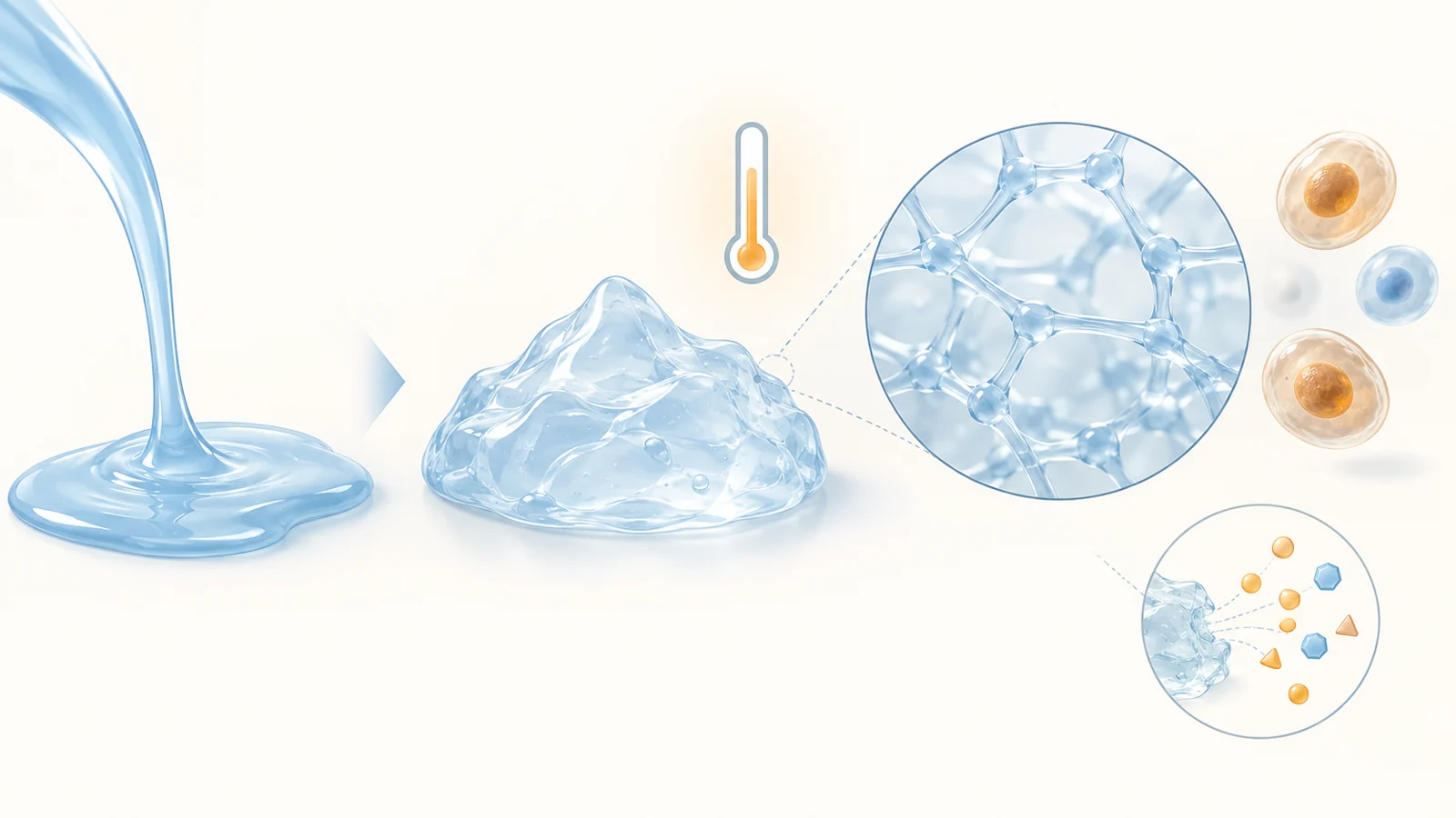
A cartilage-related example makes this tangible. Researchers used a modified chitosan formulation containing the drug kartogenin and treated with beta-glycerophosphate. At 37°C, it formed a gel within minutes. The same system was reported to sustain drug release for 40 days and enhance the development of human adipose-derived mesenchymal stem cells toward cartilage-forming cells.
Gel formation and drug release are separate behaviors on very different timescales. The temperature response changes the formulation’s physical state within minutes. Release then describes how the incorporated drug leaves that matrix over subsequent days. Combining those behaviors suggests a way to place a flowing formulation and have it become a local support under the intended conditions.
This example also helps clarify the meaning of 4D printing, another approach discussed in the review. In 4D printing, researchers print a structure using responsive materials and design it to change its properties over time when a stimulus acts on it. The design must account for the intended later change in shape, size or other properties, as well as the initial printed form.
A temperature-responsive gel does not become a 4D-printed object simply because it changes over time. The chitosan example illustrates stimulus-triggered gel formation; it does not establish that this formulation was printed. Printing adds the deliberate construction of a form whose subsequent response is part of the design.
What the different experiments actually establish
The review’s examples measure several distinct stages between making a material and repairing a tissue. Their results become easier to interpret when those stages remain separate.
Cell viability asks whether cells remain alive under the tested conditions. In one study summarized in the review, printed alginate–hydroxyapatite scaffolds produced nearly 100% viability in mouse embryo fibroblast assays after 24 and 48 hours. That is encouraging evidence about short-term compatibility with those cells. It does not show that the scaffold has formed bone or can perform bone’s mechanical work.
Other assays examine whether cells begin to display tissue-related activity. With printed scaffolds combining PLGA, a biodegradable synthetic polymer, and hydroxyapatite nanoparticles, researchers detected alkaline phosphatase after 14 days and calcium deposition after 21 days in cultures of human stem or stromal cells. These are indicators relevant to bone formation. They extend the evidence beyond survival without demonstrating a functioning replacement tissue.
Implantation tests ask a different question again. The PLGA composite study also placed scaffolds under the skin of rats and assessed the tissue response. The review reports a slight inflammatory response. Such observations help characterize how a living host responds to an implant, but a response under the skin does not establish that a material can repair a load-bearing bone defect.
An example involving a raloxifene-loaded PLGA and liquid-lipid implant goes further toward repair. The review reports sustained drug release for 55 days in release testing and well-organized bone tissue after 12 weeks of implantation in rats. The animal finding concerns newly formed tissue in a living system, giving it a different meaning from a viability percentage or a mineral-deposition measurement in culture.
None of these endpoints is interchangeable with restored human function. They concern different formulations, cell types, tissues and experimental settings, so the numbers cannot be assembled into a common performance ranking. The review is useful precisely because it brings together examples of these different questions. Its broad ambitions do not turn the featured materials into demonstrated replacements for organ transplantation or proven treatments for COVID-19 lung injury.
What this could change
For people with localized tissue defects, the prospective capability is a temporary implant that supplies physical support while delivering an ingredient intended to help repair occur at that site. In a bone defect, such a material could provide a place for new tissue to form, release a repair-promoting ingredient over time and then gradually break down as the tissue takes its place.
That possibility depends on achieving suitable mechanical function, compatibility with the host and useful repair in people. The studies gathered in this 2022 review describe components of that capability: altering cell attachment through composition, extending local drug release and using temperature to trigger gel formation. If those functions can be made to work reliably together in a clinically useful implant, a temporary support could become an active part of repairing a damaged area.
Key Sources
- Primary source: Biomaterials for Tissue Engineering Applications and Current Updates in the Field: A Comprehensive Review.
DOI or Journal Page · Peer-Reviewed Paper · 2022-09-26
Keep exploring