
Research Signal – Source date 2022-04-13
The bone surrounding a tooth helps hold it in place. Losing this support, called alveolar bone, can threaten the tooth even when the tooth itself remains. In a study published April 13, 2022, researchers tested an injectable material intended to help rebuild that supporting bone. Twelve weeks after treatment, surgically created defects in rats showed greater bone regeneration with the protein-loaded gel than with an empty gel or the untreated model comparison.
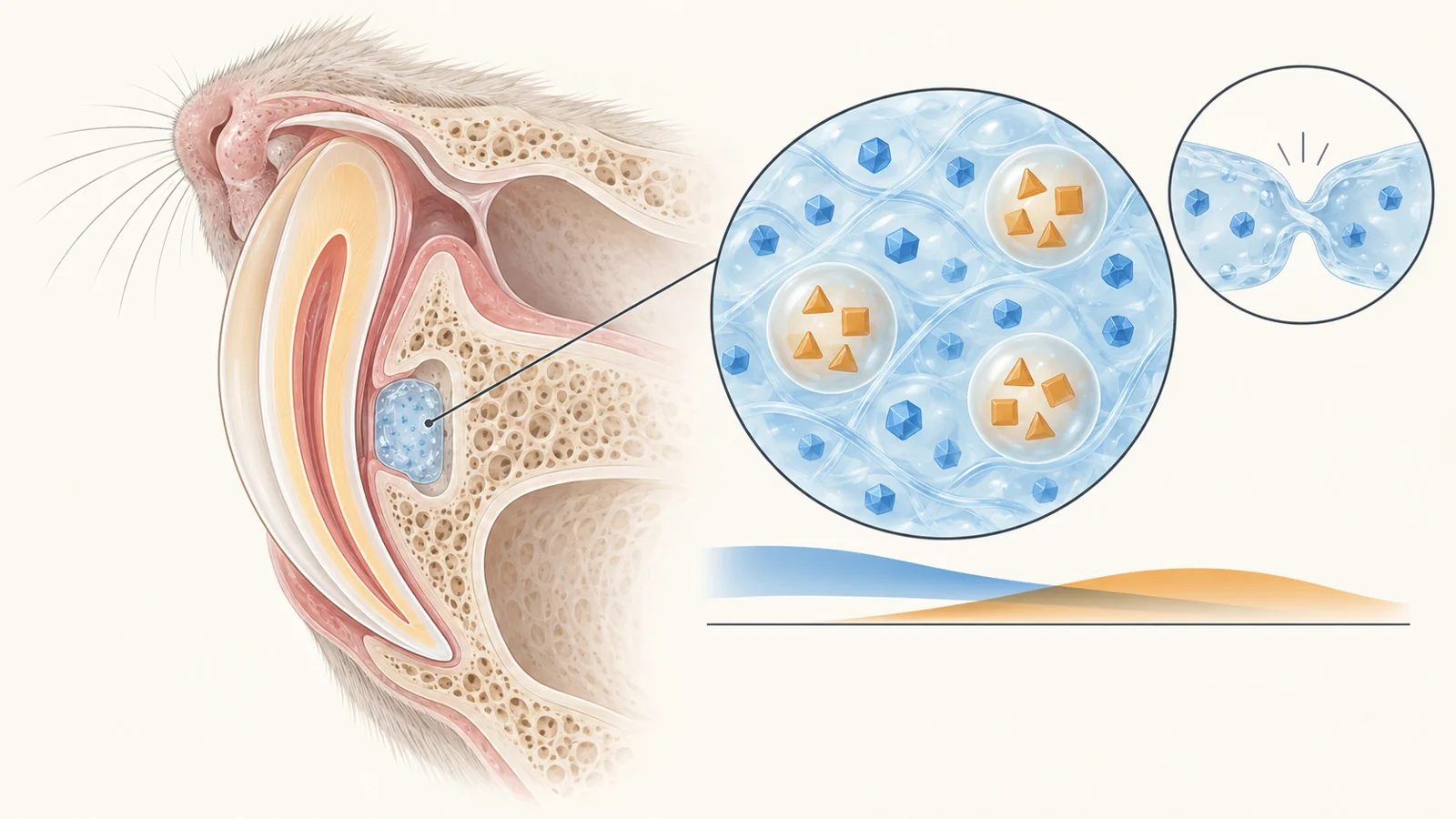
The material carries two growth-factor proteins in different compartments. One sits directly in the gel; the other is held inside tiny spheres embedded within it. That arrangement lets the proteins leave the material on different, overlapping schedules. The researchers paired this engineering work with cell experiments and animal studies to investigate whether the formulation could encourage the body’s own repair responses.
The result concerns repair of a small bone defect beside a rat tooth. Its significance lies in the combination of a material that can fit an awkward space and biological activity that translated into more new bone in that experimental setting.
A gel that can reconnect after disruption
The researchers call their formulation DFH. Its bulk material is a hydrogel—a water-rich gel—made using a chitosan-based component and a branching polyethylene glycol component. Reversible chemical links, known as Schiff base bonds, connect the network. Because these bonds can form again after disruption, separated pieces of the material can rejoin.
In a demonstration using differently colored pieces, the gel joined into a single mass after about an hour. Compression testing before and after reconnection provided further evidence that the network could recover its mechanical behavior. This is what “self-healing” means here: the material reconnects. Whether bone regenerates is a separate biological question, addressed by other experiments.
That physical behavior matters to the intended use. Bone defects in the mouth can have irregular shapes, and the available working space is narrow. An injectable, deformable material offers a way to occupy such spaces without requiring a rigid implant of exactly the right shape. The researchers therefore examined handling properties alongside swelling, degradation, and the gel’s porous structure.
Inside this network, basic fibroblast growth factor, or bFGF, is incorporated directly into the gel. Transforming growth factor beta 3, or TGFβ3, is loaded into chitosan microspheres first. Those spheres are then mixed into the surrounding material. The nested construction gives the second protein an additional compartment to leave before reaching its surroundings.
How closely the release schedule matches the biological idea
The researchers’ rationale was that different activities contribute to repair at different stages. They investigated bFGF in connection with early cell growth and blood-vessel-related behavior, and TGFβ3 in connection with cell recruitment and bone-forming differentiation. Before testing the carrier, they compared deliberately scheduled exposures in cultured human periodontal ligament stem cells—cells from the tissue that connects teeth to surrounding bone.
Those experiments included individual growth factors, simultaneous exposure, and schedules in which bFGF came first and TGFβ3 followed. Among the tested schedules, the authors reported the strongest mineralization response after approximately six days of bFGF exposure followed by TGFβ3. Staining for alkaline phosphatase, an enzyme associated with bone-forming differentiation, and for mineral deposits helped them compare the cellular responses. These were controlled culture experiments using media that supported osteogenic, or bone-forming, differentiation.
The gel produced a less sharply separated sequence. In buffered laboratory release tests, about 20% of its bFGF had emerged within 24 hours and approximately 95% by seven days. TGFβ3 remained below the assay’s detection limit during the first 24 hours, became detectable at 36 hours, and reached approximately 55% cumulative release by day 14. Its release therefore began while bFGF was still leaving the gel. The material created an early release bias followed by a longer delivery period, rather than reproducing the manually scheduled change from one protein to the other.
This distinction sets the meaning of the engineering result. The two compartments altered release behavior in buffer. Those curves do not establish the concentrations or exposure schedules that cells experienced inside a living jawbone defect, where the material interacts with tissue rather than a laboratory bath.
What the cell experiments actually observed
Several assays tested whether the formulation could support behaviors relevant to repair. Human endothelial cells, which are associated with blood vessels, formed tubular structures after 24 hours on the gel. These structures were especially numerous and distinct with the tested bFGF concentration of 80 nanograms per milliliter. The observation supports an effect on vessel-related cell organization; a tube-shaped structure in culture does not by itself establish a functioning blood supply inside repaired bone.
In a separate migration assay, cells moved through a test membrane toward the formulation. A gel containing TGFβ3 increased the number of migrating cells under the tested conditions. Periodontal ligament stem cells also survived within the gel for more than seven days, and adding TGFβ3 strengthened staining associated with bone-forming differentiation. Together, these experiments address whether cells can live in the material, move toward it, and display aspects of an osteogenic response.
The researchers also placed formulations into muscle pockets in rats and examined the gel and surrounding tissue seven days later. This was a separate implantation experiment, away from the jawbone defect. Tissue staining for the markers CD29 and CD45 was used to investigate local cellular responses. It adds an observation from living tissue, but the marker results do not trace particular recruited cells through to the new bone measured in the repair study.
The proposed mechanism thus rests on several distinct observations. Cell movement, tubular organization, differentiation-associated staining, and local tissue responses each illuminate part of the biological story. They do not amount to direct observation of one continuous sequence from recruited cells to a vascularized, rebuilt defect.
More bone in the treated rat defects
For the repair experiment, the researchers surgically exposed bone beside an upper incisor and created a defect approximately 2 millimeters across and 1 millimeter deep. Micro-CT imaging one week after surgery was used to check the defects before the treatment comparison.
The repair methods describe 24 animals assigned across four groups: the model group, a blank-hydrogel group, and two groups receiving the complete growth-factor formulation. Both loaded formulations contained bFGF at 80 nanograms per milliliter. They differed in TGFβ3 concentration, with 1,000 nanograms per milliliter in the lower-concentration formulation and 4,000 in the higher-concentration formulation. The stated injection volume was 100 microliters. These are experimental formulation details, with no established translation into a dose for people.
At 12 weeks, the loaded-gel groups had more regenerated alveolar bone than the model and blank-gel groups. Micro-CT showed improvements in three measures that describe both the quantity and internal structure of the new bone. Bone volume fraction measures how much of the analyzed region is occupied by bone. Trabecular number and trabecular thickness describe the number and thickness of the small internal bone struts.
All three measures were significantly higher in the loaded-gel groups than in the model and blank-gel controls, with the authors reporting P values below 0.05. The higher-TGFβ3 formulation performed best in the reported comparison. Tissue sections examined with standard histological stains also supported greater new bone formation in the treated defects.
These findings give the study an outcome beyond cell behavior in a dish. The complete formulation improved structural measures of repair in an animal bone injury. The blank-gel comparison is useful because it asks whether placing the carrier alone in the defect was sufficient to produce the same response; it was not in this experiment.
What the comparison can and cannot explain
The animal result supports the loaded formulation as a whole. It does not isolate how much of the benefit came from staggered release. The four specified repair groups compare two concentrations of the complete formulation with the model and empty carrier. They do not separate release order from the other effects of delivering both proteins. The scheduled cell experiments strengthen the rationale for studying timing, but they cannot supply that missing causal comparison in the rat defect.
The scale and type of injury also shape the interpretation. A surgically created hole in rat alveolar bone gives researchers a defined site in which to compare repair. It does not establish how the gel would perform in human bone loss associated with disease, or whether regenerated tissue would provide lasting support under everyday use. More bone and improved internal structure at 12 weeks are relevant findings, but they do not establish better chewing, longer tooth retention, implant survival, or treatment benefit in people. The comparisons also do not establish superiority to bone grafts or other clinical treatments.
Some reporting details limit how precisely the results can be interpreted. Although the repair methods describe 24 animals, the paper’s general statistical statement gives n = 3; these statements do not make the number contributing to each endpoint clear. The histology text also mentions groups beyond the four specified repair groups and gives differing descriptions of remaining material. Those ambiguities call for restraint in interpreting individual tissue observations and degradation, without erasing the reported micro-CT comparison.
What this could change
If the formulation can produce safe, durable repair in clinically relevant settings, dentists could gain an injectable material that fits localized defects and helps rebuild the bone supporting teeth. The intended path is local delivery of growth factors that encourage the patient’s own cells to participate in repair, potentially avoiding the need to prepare and transplant expanded cells for this approach.
For people with localized loss of tooth-supporting bone, the prospective benefit would be restoration of a foundation that helps preserve an existing tooth. The rat findings give that possibility a concrete experimental basis, while leaving lasting tooth support as a future capability to demonstrate.
Key Sources
- Primary source: Sequentially releasing self-healing hydrogel fabricated with TGFβ3-microspheres and bFGF to facilitate rat alveolar bone defect repair.
DOI or Journal Page · Peer-Reviewed Paper · 2022-04-13
Keep exploring