
Research Signal – Source date 2022-02-02
An immune treatment can affect a tumor in the spinal cord without showing that it can treat a glioma that started there. Some of the striking spinal cases discussed in a 2022 review involved cancer that had spread from elsewhere; another involved a different kind of spinal tumor. These cases matter because they suggest that immune cells and treatments can reach spinal lesions. They do not answer whether the same approach can reliably control a primary spinal cord glioma.
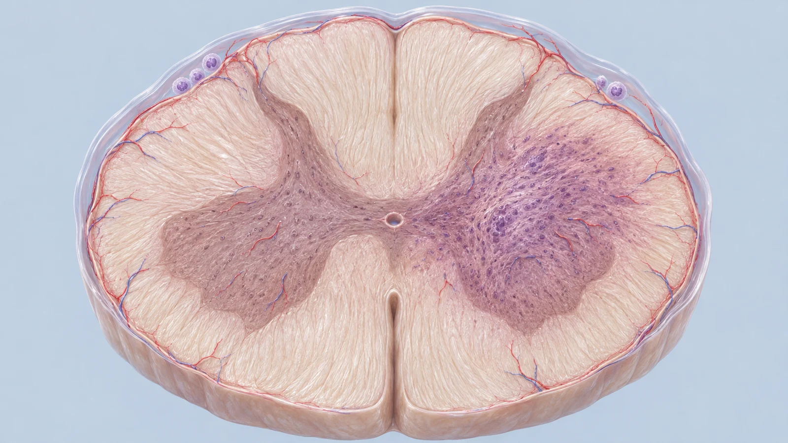
A glioma is a tumor arising from the nervous system’s supporting cells. In the spinal cord, these tumors are uncommon, and high-grade forms can grow into nearby tissue. Because the cord carries signals essential for movement and sensation, removing more tumor can mean risking lasting neurological harm. The problem is not simply finding a treatment that reaches the area. It is finding one that recognizes the right tumor and can act there without damaging the tissue around it.
A difficult tumor to study and remove
Primary spinal cord tumors account for a small fraction of tumors in the central nervous system. The review cites an incidence of about 0.22 spinal cord gliomas per 100,000 people per year. That rarity leaves individual centers with few patients and makes large comparisons between treatments difficult to organize. Much of the spinal glioma research described in the review therefore comes from small clinical reports or animal models, rather than large trials that can give a dependable estimate of benefit.
The anatomy adds a separate constraint. High-grade spinal astrocytomas can infiltrate the cord, blurring the boundary between tumor and functioning tissue. Extensive removal is often not a reasonable trade when it could cause serious neurological deficits, and the review describes standard care as centered on the safest feasible surgery, radiation, and chemotherapy. Options for more aggressive disease remain limited. This is a real reason to investigate immunotherapy, but need alone cannot tell us whether an immune treatment works.
Three ways to engage the immune system
The review groups the main strategies into checkpoint drugs, transferred T cells, and vaccines. Checkpoint drugs such as nivolumab and pembrolizumab block signals that tumors can use to restrain T cells, a type of immune cell. The aim is to release some of that restraint so the body’s own T cells can attack.
Adoptive T-cell therapies take a more direct route: T cells are collected, prepared outside the body to recognize a tumor target, and returned as treatment. A vaccine instead presents selected tumor material to prompt an immune response. These methods differ, but targeted approaches share a basic requirement: the tumor must carry a marker the treatment can recognize. Even then, that recognition has to translate into control of the disease.
Brain trial results do not settle the spinal question
The larger clinical record discussed in the review comes mainly from brain glioblastoma, a distinct setting. In CheckMate 143, nivolumab did not meet its primary goal of improving overall survival compared with bevacizumab. That result is a useful reminder that an immune mechanism or promising earlier findings do not automatically produce longer survival, even in the brain tumors studied directly.
A much smaller pembrolizumab pilot offered a more suggestive result. Among patients receiving the drug before surgery and afterward, median overall survival was 13.7 months, compared with 7.5 months for the group receiving it only after surgery. The study also found changes in tumor samples consistent with increased T-cell activity. It was a small pilot in brain glioblastoma, not a trial of primary spinal gliomas; its analysis excluded patients receiving high-dose steroids, a distinction that matters when comparing it with other checkpoint trials.
Steroids are one treatment-design complication. They may be used to manage swelling, yet the review notes concern that they can also weaken T-cell activity. In CheckMate 143, patients taking corticosteroids had worse outcomes in both treatment groups, with a larger reduction in the nivolumab group. The authors discussed steroid use as one possible contributor, alongside differences in disease severity. For spinal tumors, where inflammation itself can threaten function, treatment plans would need to account for both the immune response and the medicines used to control its effects.
Finding a target with little tissue to spare
Engineered T cells and tumor vaccines need a tumor marker that the immune system can recognize. Spinal and brain gliomas do not always share the same molecular profile, so a target that looks promising in brain tumors may not be common in spinal ones. The review notes that mutations in IDH1, often found in brain astrocytomas, appear uncommon in spinal astrocytomas. H3.3K27M is a more plausible shared target for a subset: the review reports it in 40%–50% of diffuse spinal cord astrocytic tumors. That makes it a candidate for some patients, not a universal marker for spinal gliomas.
Working out which targets are present is also constrained by the amount of tissue available. Infiltrating tumors may not have a clear surgical edge, and removing more tissue can put neurological function at risk. The review cites much smaller volumes for intramedullary spinal lesions than for brain glioblastomas, though the figures come from separate studies. Less tissue available for study makes it harder to discover targets or develop approaches that use material from the tumor. If treatment depends on a single marker, tumor cells without that marker could also persist and contribute to treatment resistance. A useful target therefore needs to be both present in the relevant tumor and characterized well enough to guide a treatment.
Getting treatment to the cord is only one part of the problem
The blood–spinal cord barrier is a protective interface between blood and spinal tissue. It regulates what passes into the cord, so it can also complicate drug delivery. The review describes evidence that this barrier may differ somewhat from the blood–brain barrier, but those differences do not make delivery to spinal tumors a solved problem. It discusses routes such as delivery into cerebrospinal fluid and the nose, as well as drug-delivery systems intended to improve access. A route that seems capable of reaching the region is a starting point, not proof of benefit in a primary spinal glioma.
Animal work also suggests that location changes how an experiment behaves. In a mouse model, CAR T cells delivered locally into cerebrospinal fluid improved survival compared with intravenous delivery in models of medulloblastoma and ependymoma. Those were not human trials of primary spinal gliomas. Other research discussed in the review found that glioma cells transplanted into brain or spinal tissue developed features influenced by where they grew. The surrounding tissue, including its immune environment, can shape what a tumor and a treatment do.
There is an added safety concern in the spinal canal: immune activity can bring inflammation and swelling, while the cord has little room to accommodate expansion. An animal study of CAR T treatment for a particular diffuse midline glioma found substantial swelling-related toxicity in a brain location where swelling was dangerous. Its spinal model did not show the same toxicity, but the tumor cells had been injected into the medulla rather than the spinal cord itself to avoid paralysis. That finding supports careful study of location-specific risks; it does not establish that treatment would be safe in people with primary spinal cord gliomas.
Spinal cases offer clues, not a single answer
The human cases help show why the origin and diagnosis of each tumor matter. The review describes immune-treatment cases involving metastatic lung cancer in the spine, a spinal melanocytic tumor treated with nivolumab, and spinal metastases from a brain glioblastoma treated with CAR T cells delivered into cerebrospinal fluid. These reports point to immune activity in spinal lesions across different diseases. They cannot be combined into evidence that a primary spinal glioma responds in the same way.
A small vaccine pilot for diffuse midline glioma included one participant with a thoracic spinal cord tumor. The review reports results for a group that also included tumors in other locations, not for that individual patient separately. The group-level findings therefore cannot tell us what happened in the spinal case. Diagnosis-specific evidence matters: a response in a metastasis, a different spinal tumor, or a mixed-location group does not supply a reliable treatment estimate for primary spinal glioma.
What this could change
A treatment matched to a marker found in a patient’s primary spinal glioma, delivered in a way that reaches the tumor and limits harm to nearby cord tissue, could eventually offer an option where current choices are few. Getting there would require more than demonstrating that immune cells can enter the spinal region. A meaningful milestone would be human evidence in clearly identified primary spinal gliomas that links the tumor’s target, the treatment delivered, and both tumor control and neurological outcomes. If that evidence holds across enough patients, clinicians could begin to distinguish who might benefit from a specific immune approach and what risks it carries. The 2022 review describes reasons to pursue that possibility, but it does not establish that capability yet.
Key Sources
- Primary source: Glioma Immunotherapy: Advances and Challenges for Spinal Cord Gliomas.
DOI or Journal Page · Review or Meta-Analysis · 2022-02-02
Keep exploring