
Research Signal – Source date 2026-02-01
A brain scan can reveal differences even when a person’s thinking appears normal. In a study of adults with moderate-to-severe obstructive sleep apnea, researchers found lower glucose uptake across frontoparietal regions and higher uptake in the cerebellum compared with a group of people without known sleep apnea. The findings raise a question: do these patterns reflect early effects of disrupted breathing during sleep, or can the study separate that possibility from other explanations?
What the PET scans measured
The researchers used positron emission tomography, or PET, with a glucose-based tracer. Glucose is sugar that cells use for energy; the scan shows where the tracer is taken up in the brain. This offers an indirect view of brain function, not a direct picture of damaged neurons or permanent injury.
The sleep-apnea group included 30 cognitively normal patients referred to a university hospital neurology unit. All had moderate-to-severe obstructive sleep apnea, defined in the study by an apnea–hypopnea index of at least 15 events per hour. Participants had sleep evaluations, cognitive testing, overnight sleep recordings, and brain imaging. The researchers excluded people with several neurological, psychiatric, sleep, and systemic conditions, as well as those with high blood pressure or signs of substantial brain changes on MRI.
For comparison, the team drew on 68 cognitively normal participants in the Alzheimer’s Disease Neuroimaging Initiative database. Sleep-apnea status in that group was screened using medical-history responses, including self-reported sleep apnea or related sleep disorders. These were database controls, rather than participants recruited and assessed through the same clinical process as the patients.
Two regions showed different uptake patterns
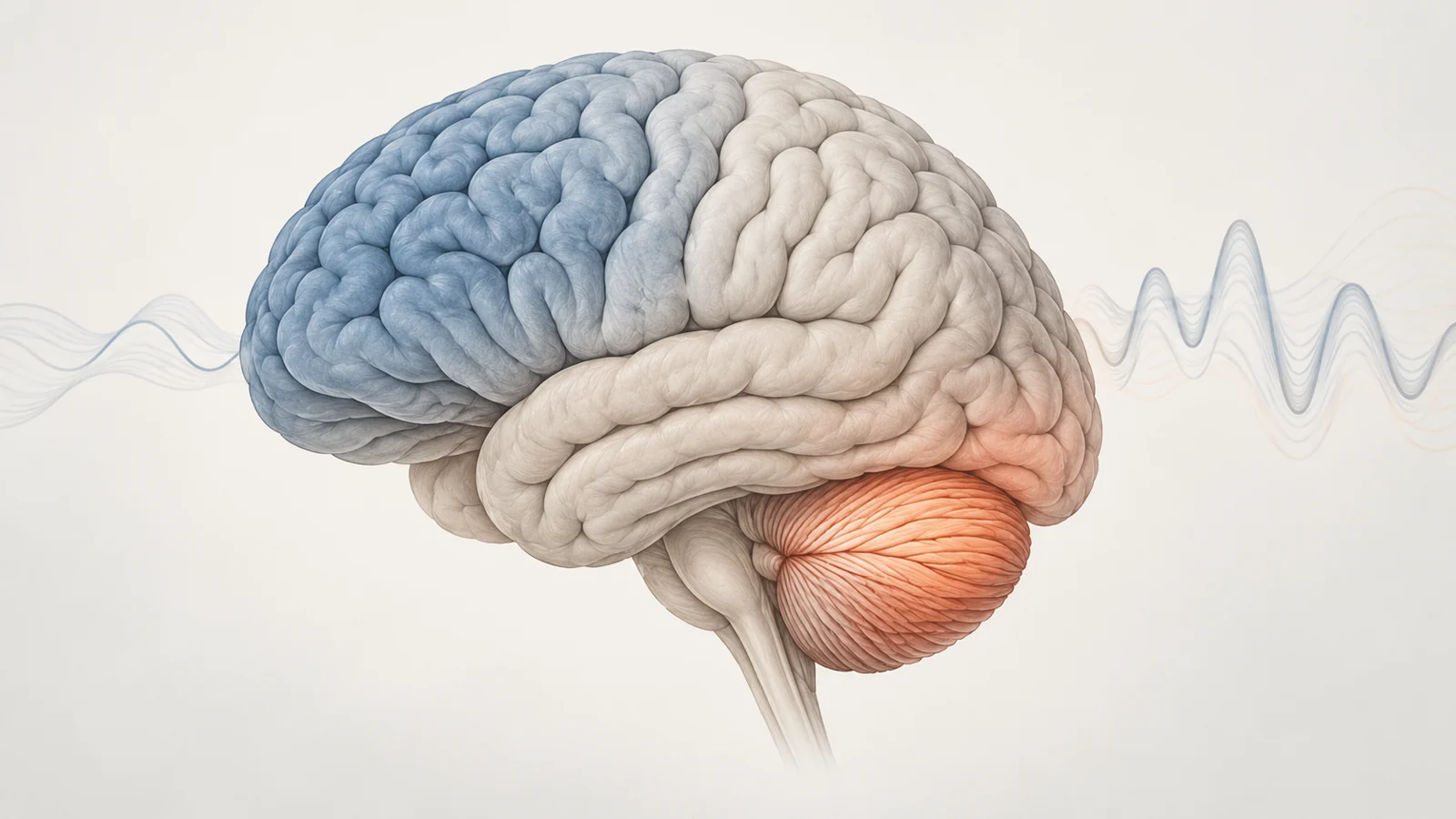
In the group comparison, glucose uptake was lower in frontoparietal areas among patients with sleep apnea. These regions include parts of the frontal and parietal cortex. A separate analysis found higher uptake in the cerebellum, a region at the back of the brain.
The researchers also examined patterns across individual patients. Their analysis found a shared low-uptake pattern involving several frontal and parietal areas, along with other regions including the hippocampus and thalamus. This was a distinct analytical step from the group-level comparison that identified both frontoparietal lower uptake and cerebellar higher uptake.
Higher uptake does not automatically mean healthier or more effective activity, just as lower uptake does not by itself establish injury. The scan registers a difference in glucose use. Its meaning depends on further evidence about how the pattern relates to sleep, cognition, and change over time.
The cerebellar finding tracked with REM sleep measures
Within the sleep-apnea group, higher cerebellar uptake was associated with shorter REM latency and a lower percentage of sleep spent in REM. REM latency is the time until REM sleep begins; the study measured both that interval and REM duration during overnight sleep recordings. The correlations describe how the measures varied together among these patients. They do not show which one drove the other.
The authors proposed that increased cerebellar activity might be a compensatory response, helping maintain functions related to REM sleep amid disrupted sleep. They also discussed other possible interpretations, including the possibility that increased activity could interfere with normal REM processes. These are explanations for the observed relationship, not mechanisms demonstrated by this comparison.
Network patterns add another layer
The team also studied metabolic connectivity: how glucose uptake in a selected seed region correlated spatially with uptake elsewhere in the brain. This method compares patterns of correlated metabolism; it is not a direct measure of communication between neurons.
Compared with controls, the patients’ patterns differed most in the attentional and limbic networks. The study assessed several large-scale networks, and the spatial overlap with a reference network template was not significant for those two. This suggests that the network patterns in those areas were less similar to the comparison patterns under the analyses used. It does not identify a specific day-to-day attention or emotional problem in an individual patient.
Why the comparison cannot show progression
The age difference complicates interpretation. The patients’ mean age was 46.87 years, while the database controls’ mean age was 72.4 years. Although the discussion describes the controls as age- and sex-matched, the reported mean ages differ substantially. The researchers included age as a covariate in some analyses, but adjustment does not make the groups equivalent in every respect. The control group’s sleep-apnea screening also relied on medical history, whereas patients underwent clinical sleep assessment.
The study captured one point in time. It cannot establish that sleep apnea caused the imaging differences, that the differences represent permanent brain injury, or that participants later developed cognitive impairment or dementia. It also did not measure whether treatment changed these participants’ PET findings. The results therefore show an association worth investigating, while leaving open whether the patterns reflect apnea-related effects, other group differences, or a mixture of factors.
What early brain changes could mean for sleep apnea care
If comparable studies confirm that these PET patterns track meaningful changes in brain function, imaging might eventually help identify people whose brains are changing before cognitive symptoms appear. Clinicians could then assess whether timely treatment of sleep apnea protects that function. This study does not validate PET for screening individuals or choosing treatment; more comparable control groups, follow-up over time, and direct treatment testing would be needed before that use could be supported.
Key Sources
- Primary source: Altered brain glucose metabolism and connectivity in young adults with obstructive sleep apnea.
DOI or Journal Page · Peer-Reviewed Paper · 2026-02-01
Keep exploring