
Research Signal – Source date 2022-09-16
A cell that stops dividing might sound inactive. Cellular senescence is more complicated. Senescent cells have entered a largely permanent state of growth arrest, but they can remain metabolically active, resist programmed cell death and release signals that change the behavior of nearby cells.
A review published on September 16, 2022, examines how this biology may connect aging and obesity with metabolic dysfunction. Its focus is not a newly completed treatment experiment, but a synthesis of cellular mechanisms, tissue observations, animal studies and early human research. The central question is whether selectively targeting senescent cells could improve how tissues handle nutrients and respond to insulin.
Why fat tissue matters for insulin sensitivity
Insulin sensitivity describes how strongly cells respond to insulin’s instructions, including signals that help regulate glucose use and storage. When tissues become less responsive, the body may need more insulin to produce the same effect. Fat tissue is not merely a passive storage depot in this process. It contains mature fat cells, immune cells and precursor cells that can produce new fat cells as tissue expands or changes.
The review describes two connected ways senescence may disrupt this system. First, preadipocytes, also called adipose progenitor cells, can become senescent. These cells show reduced capacity to replicate and develop into mature fat cells. Preadipocytes taken from obese people displayed more markers of senescence and less replicative potential than those taken from lean people. Reduced progenitor function may help explain why hypertrophic obesity is associated with enlarged fat cells and poorer tissue adaptability.
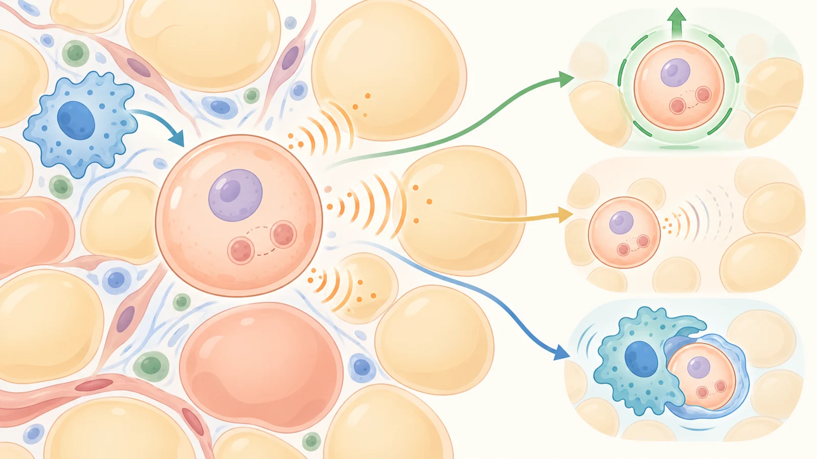
Second, senescent cells can influence their neighbors through the senescence-associated secretory phenotype, usually shortened to SASP. This is a collection of substances released by senescent cells; some can promote inflammation or alter the surrounding tissue. In adipose tissue, the review discusses factors including Activin A, interleukin-6 and tumor necrosis factor alpha. These signals can interfere with the development of neighboring precursor cells, affect insulin sensitivity and attract immune cells that may add to the local inflammatory environment.
The proposed problem is therefore broader than a few damaged cells. A senescent precursor may lose its own ability to support healthy tissue renewal while also sending signals that make nearby cells less responsive to insulin. In a condition such as obesity, those effects could reinforce one another.
Removing cells tests a causal link
An association between senescence markers and metabolic dysfunction does not by itself show that senescent cells are driving the problem. The experiments summarized in the review become more informative when researchers selectively remove cells with senescence-associated features and then measure what changes.
In obese animals, genetic approaches aimed at cells expressing high levels of p21Cip1 or p16Ink4a, as well as senolytic drugs, improved whole-body and adipose-tissue insulin sensitivity. Other measures of fat-tissue function also improved, including adipocyte size, the ability of precursor cells to form new fat cells and macrophage infiltration. These findings support the idea that senescent cells in adipose tissue can contribute to obesity-related metabolic dysfunction.
Transplantation experiments add another layer to the argument. When fat tissue from obese mice was transplanted into lean mice, removing highly p21Cip1-expressing cells from the transplanted tissue reduced the metabolic dysfunction that usually followed. In another experiment, adipose tissue from obese humans was treated with a senolytic drug before being transplanted into immunodeficient mice. The recipient mice did not develop the same insulin resistance seen after receiving untreated human adipose tissue.
The origin of that tissue matters. The fat came from humans, but the metabolic outcome was measured in mice after transplantation. This is evidence that treatment changed the behavior of transplanted tissue in an animal model, not evidence that senolytic treatment improved insulin resistance in the people who donated the tissue.
Three ways to target senescence
The review separates several strategies that act on different parts of the problem.
Senolytic drugs are intended to remove susceptible senescent cells. These cells can increase anti-apoptotic defenses, sometimes called senescence-associated anti-apoptotic pathways, that help them resist their own death signals. Senolytics inhibit those defenses, allowing selected senescent cells to undergo apoptosis. The proposed logic is selective vulnerability rather than continuous suppression of every senescent cell. The review discusses intermittent “hit and run” treatment as a way to clear susceptible cells without requiring constant exposure, but that mechanistic rationale is not a safe clinical regimen.
A second approach is to modify the SASP rather than remove the cell. Drugs described as SASP inhibitors, or senomorphic drugs, aim to reduce some of the signals that spread inflammation, alter neighboring cells or hinder immune clearance. The review discusses metformin, rapamycin and ruxolitinib in this context. Such treatments may leave the senescent cell in place while changing what it releases. Their effects can also vary by cell type, so a drug may act in a senolytic way in one setting and a senomorphic way in another.
A third strategy is to help the immune system recognize and clear senescent cells. Aging and chronic sterile inflammation can impair immune-cell function, while immune cells themselves may develop senescence-associated changes. Reducing harmful secretions could make clearance easier, and more targeted recognition strategies are also being investigated. In one mouse experiment discussed by the review, vaccination against GPNMB reduced senescent-cell burden and improved glucose tolerance. That result is an animal finding, not evidence that a comparable vaccine works in people.
These approaches leave different biological situations behind. Removal reduces the number of targeted cells. SASP suppression may keep cells present but quiet some of their effects. Immune-directed strategies depend on recognition, tissue context and the ability of immune cells to complete clearance. The distinction matters because the goal is not simply to make a tissue contain fewer cells with a particular marker.
Senescence is not automatically harmful
A universal clear-all strategy would be biologically inadequate. Growth arrest is one part of senescence, while the inflammatory secretions associated with some senescent cells are another. Not every senescent cell develops the same secretory profile. The relationship between senescence markers and inflammatory gene activity can differ between fat depots, for example.
Senescence can also serve useful functions. The review describes roles in wound healing, protection against fibrosis and cancer prevention, as well as essential functions during fetal development and possible roles in parturition. Preventing senescent-cell formation everywhere could therefore interfere with protective biology. A more selective treatment would need to distinguish harmful, persistently inflammatory cell states from senescence that is serving a normal purpose.
Pancreatic beta cells show why context matters. Aging and obesity are associated with senescence-related features in these insulin-producing cells, including p16INK4a expression, reduced proliferative capacity and SASP production. Yet the functional consequences are not uniform. In one mouse experiment, increasing p16INK4a in young animals increased insulin secretion. In another, removing cells with high p16INK4a improved beta-cell function and insulin secretion. Senolytic treatment also improved insulin secretion in a mouse model of type 1 diabetes, possibly by reducing SASP-related immune damage.
Those findings do not point to one universal prediction for insulin secretion across diabetes or across tissues. They show why a marker cannot be treated as a complete definition of a harmful cell, and why the effect of targeting senescence may depend on which cells are affected and what they are doing.
What early human findings can show
The human evidence summarized in the review is an important step, but it answers a narrower question than the animal metabolic experiments. Small open-label studies found that systemic senolytic treatment could reduce p16INK4a- and p21CIP1-expressing cells in adipose tissue and epidermis in people with idiopathic pulmonary fibrosis. A separate pilot study in that disease reported improvements in several physical-function measures after treatment.
Those observations suggest that senolytic drugs can reach human tissues and alter measurements associated with senescent cells. They do not establish that the treatment improves obesity-related insulin resistance, diabetes or long-term metabolic health. The studies involved a different disease context, and a reduction in tissue markers is not the same measurement as sustained improvement in insulin sensitivity.
Interpretation is also complicated by the biology of senescence itself. No single marker provides a universal, perfectly specific readout across every cell type and tissue. The review highlights limitations involving p16INK4a, senescence-associated beta-galactosidase and other markers, and points to the need for reliable measures that can be taken without invasive tissue sampling. Differences between tissues may eventually help researchers target disease-relevant senescent cells more precisely, but they also make broad claims harder to justify.
For metabolic disease, the next standard is therefore higher than showing that a marker changes. Randomized controlled trials and additional safety data are needed to determine whether targeting senescence produces meaningful metabolic benefits and whether those benefits outweigh risks from removing or altering cells with protective functions. The review also presents the broader healthspan idea as a geroscience hypothesis; it does not establish longer human lifespan or disease-free life.
The authors disclose related financial interests, including patents on senolytic drugs held by Mayo Clinic. They state that the research was reviewed by the Mayo Clinic Conflict of Interest Review Board and conducted under Mayo Clinic conflict-of-interest policies.
What this could change
For people with obesity-related metabolic dysfunction, a successful senescence-targeting treatment could eventually help preserve the flexibility of fat tissue and improve the body’s response to insulin. The practical route would be selective: reducing the cells or secretions that impair neighboring tissue while retaining senescence’s useful roles in protection and repair.
That possibility depends on demonstrating a meaningful metabolic benefit in people, identifying which senescent states should be targeted and establishing an acceptable safety profile. If those conditions are met, treatments directed at senescent cells or their effects could become a way to address a tissue process that contributes to metabolic dysfunction across more than one organ. The review’s evidence supports investigating that possibility, not treating it as an established therapy.
Key Sources
- Primary source: Targeting cellular senescence in metabolic disease.
DOI or Journal Page · Review or Meta-Analysis · 2022-09-16
Keep exploring