
Research Signal – Source date 2025-12-19
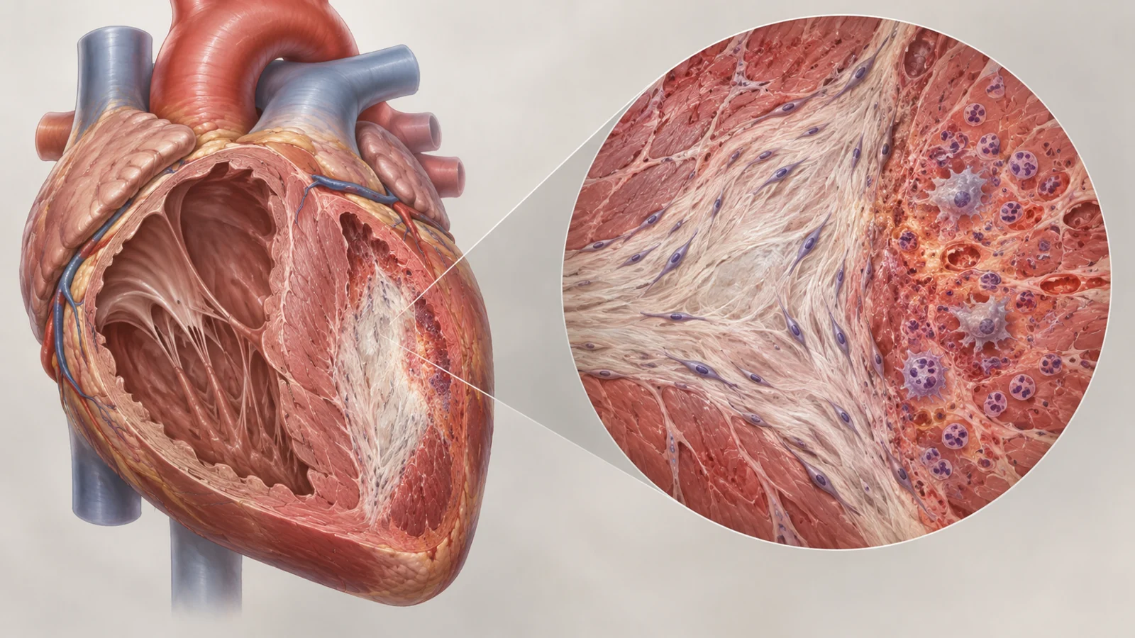
After a heart attack, removing damaged cells might seem like an obvious way to help the heart recover. But some heart cells that enter a stress-altered state may also help limit scar formation during repair. Could clearing these cells improve healing, or interrupt it?
A review published on December 19, 2025, brings together experimental work on this tension. It describes studies in which senescent cells—cells that have stopped dividing in response to stress and changed how they function—are linked both to repair-related effects and to later heart damage. The intervention results it discusses come chiefly from animal models. The review is not a new treatment trial, and its account does not establish that clearing these cells helps people recover after a heart attack.
A heart attack can push several cell types into a stress-altered state
A heart attack interrupts blood flow to part of the heart. Restoring blood flow quickly is important for patient outcomes, yet the return of blood after ischemia can add to the injury. The resulting ischemia-reperfusion injury involves reactive oxygen species, DNA damage, mitochondrial dysfunction and inflammation. In experimental models, these stresses can induce senescence-associated changes in heart muscle cells and in interstitial cells, including fibroblasts.
Senescence is not simply another name for a dead cell. It describes a stress response in which a cell stops dividing and changes its activity. Researchers track markers such as SA-β-gal, p16 and p53, but the presence of a marker alone does not tell us everything a cell is doing or what role it will play in the injured heart.
In mouse and rat infarction models, senescence markers rise over time after injury and reperfusion. Studies also report inflammatory and fibrotic signals in the heart. The findings make senescence relevant to the course of injury, but the consequences depend on which cells are affected and when.
Fibroblasts can shape the scar during repair
Fibroblasts help build and organize the scar that forms after a heart attack. In experiments summarized by the review, increasing the senescence marker p16 soon after injury improved heart function and reduced the fibrotic area. Reducing p16 weakened those effects. Infarcted mouse hearts lacking p53 had fewer senescent fibroblasts but more fibrosis and collagen deposition, another indication that fibroblast senescence can restrain scar formation in some settings.
The review also describes ATM, a kinase involved in responses to DNA damage. In experimental work, ATM-related senescence in cardiac fibroblasts was associated with less excessive collagen deposition and signals that promote blood vessel growth. ATM deficiency, by contrast, was linked to more apoptosis and fibrosis, and to worse heart function over time in mice, even though infarct sizes were comparable.
Heart muscle cells can also participate in these repair-related effects. After infarction, the GATA4 pathway can prompt release of CCN1, a secreted factor associated with protection and fibrosis regulation in experimental models. Inhibiting GATA4 increased collagen production and worsened measures of heart function in the work reviewed.
These experiments suggest that broadly eliminating every cell with senescence-associated features could interfere with processes that help organize repair. They do not identify a cell type that should always be spared or establish a treatment window for people.
Persistent senescent cells are linked to later remodeling
The concern shifts when senescent cells accumulate or persist. The review describes evidence that their secretions can become more inflammatory over time and contribute to chronic inflammation, fibrosis and adverse remodeling. In mouse models, senolytic drugs—agents intended to eliminate senescent cells—reduced some signs of hypertrophy and improved measures of cardiac function. These findings concern experimental animals, and they address a different setting from experiments in which fibroblast senescence helped limit scarring during repair.
In aged mice, studies of several senolytics found fewer senescent cells alongside improved fibrosis measures and ejection fraction. Other work linked senescent fibroblast activity to longer-term tissue damage. The review also discusses animal experiments in which dasatinib plus quercetin cleared senescent cardiac progenitor cells and improved heart measures, as well as studies in which navitoclax reduced senescence-associated markers and improved cardiac output, ejection fraction or stroke volume.
Such results make cell removal a research possibility, not a settled prescription. The effects depend on the cell populations affected and the stage of injury. The experiments do not establish a universally safe time to intervene or show that a senolytic prevents heart failure in people after a heart attack.
Inflammation measurements need a source
An inflamed heart after a heart attack does not automatically mean senescent cells produced the measured inflammatory molecules. The injury itself triggers sterile inflammation, with rapid increases in cytokines such as IL-1β, TNF-α and IL-6. Senescent cells can also release a mixture of signals, often called the senescence-associated secretory phenotype, or SASP. Some factors associated with this secretory pattern have been observed after infarction, but many inflammatory signals can arise from more than one process.
That makes attribution difficult. A rise in a cytokine, chemokine or other inflammatory marker cannot by itself identify senescent cells as its source. Understanding which cells released a signal, and at what stage of injury, matters when interpreting experiments or considering a treatment aimed at senescence.
Removing cells and changing their secretions are different strategies
Senolytics aim to eliminate senescent cells. Senomorphics instead aim to alter their behavior or suppress harmful parts of their secretory activity. The distinction matters because cell removal may also eliminate cells that are contributing to repair, while changing a secreted signal could in principle affect a narrower part of the response. The review presents both approaches as areas for investigation; it does not establish which would be safer or more effective after a heart attack.
Navitoclax illustrates the practical challenge of selectivity. In rodent infarction models, treatment was associated with better heart-function measures and changes in scar size, blood vessel growth and senescence-associated signals. But the review notes systemic toxicity concerns from cancer treatment, including severe and unpredictable reductions in neutrophils and platelets. Nanoparticle delivery of navitoclax was developed and tested in experimental ischemia-reperfusion models, with reported heart benefits and no apparent systemic side effects in those models. That is an experimental delivery approach, not evidence of a safe human treatment.
Timing remains an open question. A treatment given during an early repair response could affect a different set of cells and processes than one aimed at cells that persist later. The review does not resolve when treatment should occur, which cells it should target, or how to preserve useful repair activity while reducing harmful effects.
Human observations show relevance, not treatment benefit
The review cites human observations consistent with senescence-related changes in ischemic heart disease. Postmortem ischemic heart tissue showed increased levels of markers including p16 and SA-β-gal. Blood samples from people with ischemic heart disease showed higher levels of several SASP-associated chemokines, and bone marrow progenitor cells had shorter telomeres, more senescence markers and reduced myeloid differentiation capacity.
These observations connect the experimental subject to human disease, but they do not show that clearing senescent cells improves recovery. Tissue and blood markers do not establish which cells produced a signal, whether a particular treatment would help, or what its risks would be after a heart attack.
The review also discusses conventional cardiovascular and metabolic drugs, including metformin, losartan and levosimendan, alongside experimental changes in senescence markers. Those drugs have clinical uses or reported clinical benefits in their respective settings, but marker changes in animal models do not prove that direct targeting of cardiac senescence caused those benefits. Improved blood pressure, glucose control, oxidative stress or other systemic factors could also influence the markers.
What a better timed treatment could mean for heart recovery
A treatment that selectively reduces harmful, persistent senescent-cell activity while preserving repair could eventually improve recovery after a heart attack and reduce damaging remodeling. Reaching that possibility depends on learning which cells to target and when, and on showing in people that the benefits outweigh the risks. The studies summarized in this December 2025 review offer experimental clues, but they do not yet demonstrate that outcome for patients.
Key Sources
- Primary source: The Yin and Yang of cellular senescence in ischemia-induced myocardial injury.
DOI or Journal Page · Review or Meta-Analysis · 2025-12-19
Keep exploring