
Research Signal – Source date 2022-10-27
Aortic stenosis is a narrowing of the valve through which blood leaves the heart. It becomes more common with age, but aging alone does not explain who develops significant disease. A review published on October 27, 2022, describes a more complicated picture: age-related changes can combine with mechanical, metabolic and inflammatory stresses, while many older people never develop important valve narrowing.
The review’s central question is therefore not simply how to treat an old valve. It is whether active biological processes begin early enough that they could eventually be slowed, and whether damaged or senescent cells might become part of that treatment strategy.
Aging is only part of the explanation
Cellular senescence is a state in which a cell permanently stops dividing. It is different from quiescence, a reversible pause that allows a cell to respond to its nutrient environment. A senescent cell has lost its ability to replicate, but it is not necessarily silent. It can continue releasing cytokines, chemokines, growth factors and matrix metalloproteinases. These substances can influence nearby cells and alter the surrounding extracellular matrix.
Senescence can arise through ordinary replicative aging, including progressive telomere shortening, or through stress that damages DNA, mitochondria and other cell systems. The review connects this stress-induced form to excessive mechanical strain, oxidative stress and metabolic pressures. Chronological aging and these local stresses can overlap, but they are not interchangeable. A valve exposed to years of unusual force may experience a different biological burden from one that has simply aged.
The effect on the valve may be twofold. Senescent endothelial cells at the valve surface no longer divide to replace damaged cells, while their secretions can promote inflammation and tissue remodeling. The immune system may help remove some age-related senescent cells, but persistent stress and age-related changes in immune function can allow them to accumulate. The review also describes evidence that circulating endothelial progenitor cells become less available and more senescent with age, reducing another route for repairing a disrupted surface.
How a surface injury can become a stiff valve
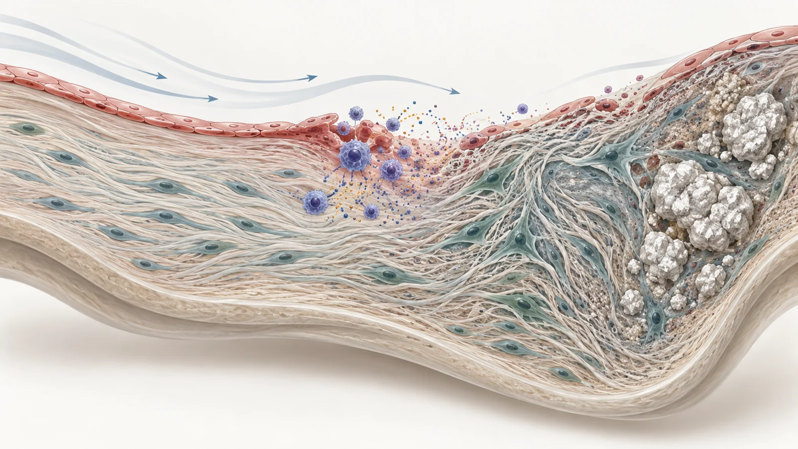
The early process often begins at the aortic side of the valve, where blood flow creates oscillating and turbulent forces. The ventricular side normally experiences more linear, high-shear flow. Mechanical conditions on the aortic side can make the endothelial barrier more vulnerable to dysfunction and focal disruption.
Once that barrier is compromised, lipids from the blood can enter the tissue beneath it. Oxidation and inflammation follow, with macrophages and other inflammatory activity contributing to remodeling. Fibrosis adds collagen and stiffness. At this stage, the valve is not simply wearing out as an inert structure. Cells and signaling systems are responding to injury and changing the material around them.
The review separates this initiation phase from a later progression phase. Valvular interstitial cells, which help maintain the leaflet, can first take on a myofibroblast-like state associated with fibrosis. Later, some acquire osteogenic, or bone-forming, characteristics. Pathways involving NOTCH and the RANK, RANKL and OPG system are among those discussed in the review as part of this shift.
Mineral deposits then add their own mechanical consequences. A stiffening leaflet opens and closes less effectively, and the altered forces can create further injury. The result is a feedback loop in which inflammation, tissue remodeling, mineral deposition and mechanical stress reinforce one another. Early lesions are not destiny: the review notes that roughly 10–15% of people with aortic valve sclerosis progress to obstructive calcification during their lifetime. Sclerosis and stenosis therefore describe different stages, and an early calcific focus should not be treated as proof that severe obstruction will follow.
Why artery treatments do not translate neatly
Aortic valve disease and arterial atherosclerosis share several early features. Both can involve endothelial dysfunction, lipid entry, inflammatory cells and microcalcification. That overlap helps explain why treatments successful in vascular disease have attracted interest in valve disease. It does not guarantee that the later biology, or the response to treatment, will be the same.
The review describes important differences. In arteries, lipids can move deeper into the vessel wall and contribute to foam cells, necrotic cores and a fibrous cap. Plaque rupture can trigger an acute event. In the valve, lipids remain closer to the subendothelial layer, and later progression is more strongly shaped by mechanical stress, fibrosis and the interaction between inflammatory cells and calcification pathways. The review characterizes valve calcification as a stable, progressive process with lamellar bone formation rather than the rupture-prone pattern associated with an unstable arterial plaque.
The treatment record summarized in the review reflects that divergence. The ASTRONOMER and SALTIRE trials tested intensive cholesterol lowering, while the SEAS trial examined simvastatin combined with ezetimibe. These studies did not show that cholesterol lowering slowed the progression of aortic stenosis. SEAS did reduce ischemic events, but the valve itself did not narrow more slowly over the follow-up described by the review.
Those findings separate two questions that are easy to combine. A therapy may reduce vascular risk without changing the biological progression of a calcifying valve. The review discusses treatment timing as one possible explanation for why lipid and inflammatory processes may be more important at initiation than later, when mechanical and osteogenic feedback has become dominant. That remains a hypothesis about disease biology, not proof that treating the valve earlier would succeed.
Diagnosis sees function later than biology begins
Transthoracic echocardiography is the gold-standard method used to assess aortic stenosis. It can show thickened, calcified cusps, reduced leaflet movement, accelerated blood flow and the pressure difference created as the narrowed valve obstructs blood leaving the left ventricle. In adults, the normal opening area is described in the review as about 3–4 square centimeters; severe stenosis is generally associated with an area of 1 square centimeter or less.
The challenge is that the biological sequence begins before the opening area changes substantially. Endothelial disruption, lipid deposition, inflammation and microscopic mineralization may be active while the valve still opens well enough that routine functional measurements reveal little. Even severe stenosis can remain without symptoms for a long period, according to the review.
This creates a gap between diagnosing established obstruction and finding an early opportunity to intervene. The review discusses computed tomography and cardiac magnetic resonance as useful additional imaging approaches, but identifies the detection of early valve disease as an unresolved clinical problem. Its 2022 perspective does not establish a validated screening program or justify routine screening of people without symptoms. It points instead to the need for more sensitive molecular or imaging tools that could identify potentially modifiable disease before later calcification becomes difficult to reverse.
Senolytics remain a future possibility
Senolytics are treatments intended to remove senescent cells by exploiting the pathways that help those cells resist apoptosis, or programmed cell death. The proposed logic in the review is straightforward: reducing a population that releases inflammatory and matrix-remodeling signals might lessen the bystander effects on neighboring cells, while also improving the tissue’s ability to repair itself.
The review discusses senescent-cell removal as a possible strategy for aortic valve disease and refers to compounds such as dasatinib, quercetin and navitoclax in the wider senolytic literature. It also notes findings from broader cardiovascular research suggesting that eliminating senescent cells may delay some disease processes. Those observations help establish a direction for investigation, but they are not clinical evidence that senolytics slow aortic stenosis.
Several uncertainties remain especially important for the valve. Researchers would need to identify which senescent cells are relevant, determine when removal could help, and show that the intervention does not damage cells needed for normal tissue maintenance. The review emphasizes that human applications remain limited by incomplete knowledge of the basic biology of senescence. It also does not establish that a future drug could replace a valve-replacement procedure when severe obstruction already requires intervention.
The practical promise therefore lies earlier in the chain. If senescent-cell signaling, failed endothelial repair or the transition toward osteogenic activity can be modified safely in people, the disease might become more treatable before calcification and mechanical feedback have advanced too far. The review presents that possibility as a direction for future work, not as an available therapy.
What slowing valve damage could mean for patients
Preserving valve function for longer could delay the need for surgical or transcatheter valve replacement and reduce the growing burden of these procedures as the population ages. That benefit would depend on a treatment safely slowing valve disease in people and reaching them while the biological process remains modifiable. Senescent-cell removal is one proposed route toward that outcome, not an established means of achieving it.
Key Sources
- Primary source: Cellular Senescence, Aging and Non-Aging Processes in Calcified Aortic Valve Stenosis: From Bench-Side to Bedside.
DOI or Journal Page · Review or Meta-Analysis · 2022-10-27
Keep exploring