
Research Signal – Source date 2025-10-02
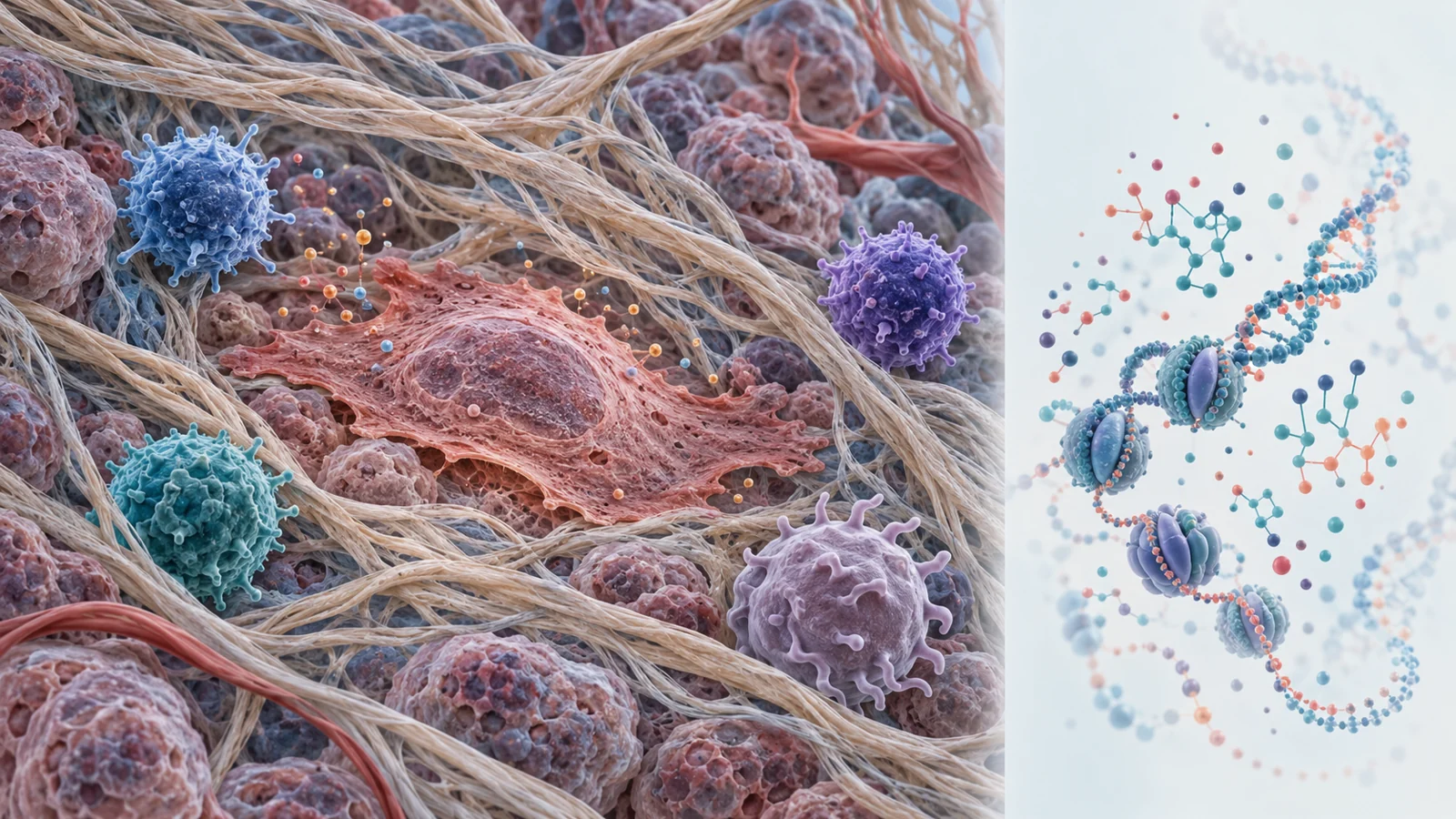
An October 2, 2025 review examines a difficult contradiction in cancer biology. Cellular senescence can prevent damaged cells from multiplying, yet persistent senescent cells can also help an established tumor avoid immune attack. The difference lies in what happens after growth stops. Senescent cells do not simply become inert. They can release signaling molecules that change nearby immune cells, cancer-associated tissue, and the extracellular matrix.
The review connects this biology with aging clocks, which estimate biological aging from molecular patterns. It also discusses selective approaches for removing harmful senescent cells and the possibility of using aging measurements to guide immunotherapy. That possibility remains a research direction rather than an established way to choose cancer treatment.
The same process can protect tissue and aid a tumor
Cellular senescence is commonly defined by a durable arrest of cell division. Two major regulatory routes highlighted in the review are the p53–p21CIP1 pathway and the p16INK4A–Rb pathway. In this state, a cell is no longer multiplying normally, which can help stop a damaged cell from progressing toward malignancy.
Senescence also involves a changed secretory profile known as the senescence-associated secretory phenotype, or SASP. These secretions can support the removal of newly damaged or abnormal cells by attracting immune effectors. In an acute setting, that response may contribute to tissue protection.
The situation changes when senescent cells persist. Their continuing secretions can produce chronic inflammation and reshape the tissue around a tumor. In the review’s account, the same broad biological program therefore has different consequences depending on timing, cell type, and local environment. Removing every cell that shows a senescence-related feature would risk confusing a protective response with a harmful, persistent population.
How a local alarm becomes an immune shelter
Inflammation is not automatically an effective attack on cancer. A persistent senescent cell population can release factors such as IL-6, CCL2, and TGF-β. These signals recruit myeloid-derived suppressor cells and M2-polarized macrophages, which can restrain effector T-cell proliferation and cytotoxicity. The tissue becomes inflamed, but the recruited immune composition may favor suppression rather than tumor clearance.
Therapy-induced senescent cancer-associated fibroblasts add another layer. The review describes these stromal cells releasing CXCL12, a signal associated with cancer stem-cell dormancy and recruitment of regulatory T cells. Regulatory T cells can help establish an immune-privileged neighborhood in which malignant cells are harder for attacking lymphocytes to reach or eliminate.
Senescent cells may also alter the recognition signals that immune cells use. The review describes reduced expression of NKG2D ligands such as MICA and MICB, which normally help activate natural killer cells. At the same time, senescent stromal cells may increase PD-L1 through p16INK4A-dependent NF-κB activity, strengthening an inhibitory signal for T cells. In simple terms, the local environment can turn down the alarm while increasing the brakes.
Physical structure matters as well. Tumor-associated extracellular matrix contains proteins such as collagen, fibronectin, laminin, and proteoglycans. When their organization or crosslinking changes, the matrix can influence blood-vessel formation, tumor-cell movement, and T-cell infiltration. An immune cell may therefore face both biochemical suppression and a remodeled tissue landscape that makes access more difficult.
An aging clock measures molecular age
An aging clock is a statistical model trained on molecular data to estimate age or the rate of aging. The estimate can be compared with chronological age to identify a difference between the two. DNA methylation clocks are the most established example discussed in the review. They use patterns of methyl groups attached to DNA at CpG sites, where those patterns can be associated with changes in gene regulation.
The review notes the development of clocks including Horvath’s and Hannum’s models, as well as later measures such as DNAmPhenoAge and GrimAge. These tools are intended to capture molecular features associated with aging and, in some cases, age-related disease risk or survival. They can provide a quantitative way to study whether an intervention changes an aging-related measurement.
An aging-clock estimate is not the same thing as locating or counting senescent cells inside a patient’s tumor. It summarizes a molecular pattern from a particular sample. The number may be useful for studying biological age, but it does not by itself identify which cells are senescent, where those cells are, or whether they are helping or hindering immune treatment.
The measurement is not a tumor census
A clock trained in one tissue may not work equally well in another. The review describes relatively low agreement between epigenetic age estimates from blood and oral tissue, with bias appearing when a blood-derived model is applied to oral samples. Differences in cellular composition and tissue-specific epigenetic features can change what a molecular age estimate means.
This issue matters for cancer research because a blood measurement may reflect systemic or immune-related biology without directly representing the tumor microenvironment. A tissue-appropriate measurement could be more informative, but the review presents cross-tissue application as an unresolved analytical challenge rather than a settled clinical practice.
Cell identity creates a second measurement problem. Markers often associated with immune-cell senescence, including loss of CD28 or increased CD57 and KLRG1, can also appear in other dysfunctional or highly differentiated states. CD57, for example, can mark terminally differentiated, highly cytotoxic natural killer cells without proving that those cells are senescent. A marker panel therefore needs context; one marker cannot settle the biological identity of an immune cell.
Selective delivery is still a preclinical idea
The therapeutic concept described by the review is selective intervention. Instead of broadly suppressing aging-related biology, researchers could try to eliminate senescent stromal or tumor-associated cells that are sustaining an unhealthy environment, or reduce the harmful parts of their secretory program while preserving useful functions.
One example gives that idea physical detail. The review describes roughly 100-nanometer silica nanoparticles coated with six-mer β(1,4)-galacto-oligosaccharides and loaded with doxorubicin or navitoclax. Senescent cells in the cited work had about two to three times higher senescence-associated β-galactosidase activity. That enzyme activity could break down the particle coating and release the drug near the targeted cells.
In palbociclib-treated SK-MEL-103 melanoma xenografts, the doxorubicin-loaded particles reduced tumor volume by 47%, compared with 22% for free doxorubicin in the model summarized by the review. The same report described reduced cardiotoxicity with the targeted formulation. These are results from a preclinical xenograft experiment, not evidence of human treatment benefit, survival improvement, or general safety.
The example is important because it shows what “selective” might mean in practice: a delivery system designed around a feature enriched in senescent cells rather than an assumption that every aging-related cell should be removed. It also shows why the route to clinical use is demanding. The relevant enzyme activity, tissue distribution, drug exposure, and safety profile would all need to remain useful in people.
Where the clinical promise narrows
The review proposes that aging-related biomarkers could eventually help researchers distinguish patients whose immune and tissue environments are more likely to respond to particular immunotherapies. It also discusses interventions studied in human settings, including work involving rapamycin, everolimus, immune-cell phenotypes, and vaccine responses in older adults.
Those findings address different populations and endpoints from cancer treatment. A change in antibody response or an immune-cell measurement in a non-cancer study does not demonstrate that assigning a checkpoint inhibitor or another cancer immunotherapy according to an aging-clock reading improves tumor control. The review’s proposal depends on connecting a molecular measurement to a treatment decision and then showing that the decision improves outcomes.
Several obstacles remain before that connection could be trusted. The review points to small samples in early clinical research, limited longitudinal safety information, and the difficulty of representing aging as a multidimensional biological process. Measurements would also need to be validated in the tissues and immune contexts where they are meant to guide care. A clock can be statistically accurate about an age-related pattern while still being clinically unhelpful for choosing a particular therapy.
What this could mean for older cancer patients
From the review’s October 2025 perspective, the longer-term possibility is more individualized immunotherapy that considers the patient’s immune and tissue environment alongside the tumor itself. Older patients could eventually benefit from combinations that address harmful senescent populations, suppressive signals, or barriers created by the surrounding matrix while preserving immune functions that help control abnormal cells.
That future depends on more than producing a precise-looking biological-age number. Tissue-relevant biomarkers would need to provide useful treatment guidance, selective interventions would need to show clinical benefit, and both would need acceptable long-term safety. If those conditions are met, senescence biology and aging clocks could add information about a patient’s immune and tissue environment to cancer-treatment decisions. At the publication date, that remains a conditional possibility grounded in a review of molecular, preclinical, and human research with different levels of evidence.
Key Sources
- Primary source: Reprogramming cellular senescence and aging clocks for advanced cancer immunotherapy.
DOI or Journal Page · Review or Meta-Analysis · 2025-10-02
Keep exploring