
Research Signal – Source date 2022-02-28
Aging bones become clinically important when reduced strength leads to fractures. Hip, wrist, and vertebral fractures can cause pain, disability, deformity, and increased mortality. Osteoporosis develops through several routes. After menopause, estrogen deficiency is a major driver, while age-related osteoporosis also involves inflammatory processes, calcium and vitamin D insufficiency, higher parathyroid hormone levels, and impaired activity of bone-forming cells.
A February 2022 narrative review asks whether another feature of aging could be part of this process: the accumulation of senescent cells. These cells have stopped dividing, but they remain metabolically active and can alter nearby tissue. The idea is not to replace established osteoporosis treatment, but to investigate whether addressing senescence could eventually add another way to protect the skeleton.
Why aging bones become fragile
Bone is continually renewed through remodeling. Osteoclasts remove older or damaged bone, while osteoblasts produce new organic matrix and coordinate its mineralization. The two cell types work in a linked cycle that helps preserve mechanical strength and maintain calcium and phosphate balance. Resorption takes roughly two to four weeks, while the subsequent formation process takes about four to six months.
When removal and formation become unbalanced, bone mass and microarchitecture can deteriorate. Osteoporosis is defined by low bone mineral density together with a broader impairment of bone structure that increases fracture risk. The remodeling problem is therefore not simply that bone disappears. The quality and organization of the remaining framework also matter.
The review places senescence-directed strategies alongside existing osteoporosis-specific medicines. Antiresorptive drugs have reduced vertebral, non-vertebral, and hip fractures in older adults in the evidence summarized by the authors, and anabolic treatments and other bone-specific options are also part of the treatment landscape. Targeting senescence remains a proposed addition to this landscape, not an established replacement or superior approach.
Senescence changes the bone neighborhood
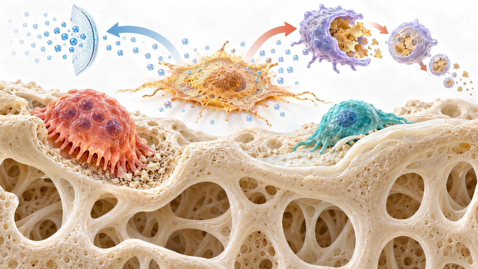
Quiescent cells can stop dividing temporarily and later re-enter the cell cycle when they receive the right signals. Senescent cells generally remain in a more permanent non-proliferative state. They also become larger and show increased metabolic and lysosomal activity. Their most important effect for this discussion may be the senescence-associated secretory phenotype, or SASP.
The SASP is a mixture of secreted molecules that can include inflammatory cytokines, chemokines, growth regulators, angiogenic factors, and matrix-modifying enzymes. These signals can influence neighboring cells in different ways depending on the tissue and circumstances. Senescence is therefore not just a label attached to an inactive cell. It describes a change in the local signaling environment.
Evidence summarized in the review connects this environment with age-related bone loss. In aging rats and mice, proinflammatory and senescent immune-cell subtypes accumulated in bone marrow and secreted granalcin. The review describes this signal as repressing osteogenesis, the process of producing bone, while promoting adipogenesis, the formation of fat cells. A shift in the marrow environment away from bone formation offers one possible route by which cellular aging could affect the balance between osteoblasts and osteoclasts.
Other findings described by the review suggest communication between bone and non-bone tissues. In one line of work, extracellular vesicles from mouse and human muscle cells with elevated levels of a molecule called miR-34a induced senescence in mouse bone-marrow stromal cells. Vesicles from senescent osteoblasts also affected endothelial-cell function. These observations fit with the close relationship between blood-vessel formation and bone formation, but they remain pieces of a biological explanation rather than a clinical treatment result.
What mouse experiments add
Observing senescence-associated changes is different from showing that they cause bone loss. The review reports both kinds of evidence. Senescence markers increased in several bone-related cell populations from older mice, including osteoprogenitors, osteoblasts, osteocytes, and immune cells. Older postmenopausal women also had more senescent cells in bone biopsy samples than younger premenopausal women in the comparison described by the review.
The human biopsy observation is important because it shows that the biology is not limited to laboratory animals. It does not, however, show that clearing those cells improves human bone outcomes. A tissue comparison can identify an association between age and senescence without establishing that senescent cells are the intervention point.
The review describes stronger causal evidence from old mice. Pharmacological clearance of senescent cells, as well as genetic elimination of cells expressing the senescence-associated marker p16Ink4a in a specialized mouse model, improved bone microarchitecture and bone strength. The experiments also showed increased bone formation and reduced bone-marrow fat. The authors relate these effects partly to the removal of senescent osteocytes and to a shift in the behavior of marrow-derived mesenchymal stem cells away from adipocytes and toward osteoblasts.
These results make senescent cells a credible target for further study because an intervention changed skeletal outcomes in animals. They still answer a narrower question than whether a senolytic treatment prevents fractures in people with age-related osteoporosis. Mouse bone strength and architecture are valuable experimental outcomes, but they are not clinical fracture-prevention results.
Two ways researchers might intervene
The review separates two treatment ideas. Senostatics, also called senomorphics, aim to reduce the harmful components of the SASP while leaving the senescent cells in place. Senolytics are drugs intended to promote the selective death of senescent cells. The first strategy changes the signals released into the tissue; the second changes which cells remain there.
The exposure patterns could differ. Suppressing a secretory pathway may require continuing treatment, but SASP molecules also have functions unrelated to senescence, so prolonged inhibition could affect other biology. The review uses ruxolitinib as a preclinical example: blocking the Janus kinase pathway reduced multiple SASP components and improved bone microarchitecture and strength in 22-month-old mice. That finding illustrates the secretion-suppression route without establishing it as the best strategy.
Cell-clearing treatment may instead be given intermittently. Senolytic compounds are intended to weaken the survival pathways that allow senescent cells to resist the inflammatory and pro-apoptotic pressures they generate, making temporary exposure a possible approach. The distinction matters because the treatment is acting on living cells with roles in both harmful and beneficial processes. A shorter dosing schedule may change risk, but it does not remove the need to establish which cells are affected and what happens afterward.
What the early human evidence measured
The review describes human evidence at two different stages. Preliminary findings from people with diabetic kidney disease involved an oral combination of dasatinib and quercetin given for three days. The reported changes included lower blood levels of some SASP components and fewer senescent cells. Those findings concern senescence-associated measures in another disease population. They do not demonstrate improved bone strength or clinical benefit for osteoporosis.
The review also describes a registered open-label phase 2 study designed around skeletal health in 120 older women. The planned groups were to receive intermittent dasatinib plus quercetin, intermittent fisetin, or no intervention. The stated primary outcomes were changes over 20 weeks in CTX, a marker of bone resorption, and P1NP, a marker of bone formation. At the time of the review, results were not yet available.
Those measurements could show whether treatment changes bone turnover markers. They would not, by themselves, directly demonstrate fewer hip, wrist, or vertebral fractures. Fracture prevention is the clinical outcome that matters to patients, but the trial description in this review does not provide that outcome. The animal findings, biopsy comparisons, diabetic-kidney-disease results, and planned skeletal-health study therefore belong to separate levels of evidence and should not be combined into one demonstrated benefit.
Safety depends on what senescent cells do
A senescent cell can contribute to disease-related signaling, but senescence also has physiological roles. The review notes functions that include tumor suppression and participation in tissue repair, including wound healing. Clearing these cells or suppressing their secretions could therefore produce effects that depend on tissue, timing, dose, and the particular drug.
The review gives concrete examples. Navitoclax was associated with platelet toxicity, including transient thrombocytopenia and impaired platelet function. High concentrations of quercetin inhibited osteoblast differentiation in cell experiments, raising a bone-related concern about excessive exposure. In aged mice, continuous navitoclax treatment at 50 mg per kilogram per day for two weeks caused trabecular bone loss, whereas the broader senolytic idea often contemplates intermittent dosing.
The same evidence set also complicates the assumption that senescent-cell clearance must impair repair. After femoral fractures, dasatinib plus quercetin cleared senescent cells and reduced SASP markers in the fracture callus without impairing healing; the review reports that healing was even accelerated in that experiment. A related rat study found increased bone regeneration in calvarial defects under conditions intended to increase stress-induced premature senescence. These findings do not prove safety in people, but they show why the biological question cannot be reduced to “remove every senescent cell.”
A useful treatment would need to distinguish damaging signaling from necessary repair, and harmful cell accumulation from beneficial transient senescence. It would also need a regimen that changes skeletal biology without causing platelet problems, disrupting osteoblast activity, or producing bone loss through excessive or continuous exposure.
How targeting senescent cells could help protect aging bones
Targeting cellular senescence could eventually offer an additional osteoporosis treatment aimed at an aging-related disturbance of bone renewal. The potential benefit is concrete: stronger bones could reduce the likelihood that an ordinary fall or other stress becomes a disabling fracture. Reaching that outcome would require human evidence that the treatment improves meaningful skeletal health, followed by evidence that the benefit outweighs toxicity and preserves necessary repair functions.
The February 2022 review supports the biological plausibility of this direction. It connects senescent-cell accumulation, inflammatory signaling, marrow-cell behavior, and altered bone remodeling; it also summarizes animal interventions in which clearing senescent cells improved bone architecture and strength. The human observations and treatment studies described there had not yet established fracture prevention in people with age-related osteoporosis.
For readers, the important question is therefore not whether senolytics are already a proven answer. It is whether carefully targeted, appropriately timed control of senescent cells can produce meaningful human skeletal benefits without damaging the systems that maintain blood cells, bone formation, and tissue repair. Protecting aging bones remains the goal, and the route from promising biology to fewer fractures still depends on that clinical test.
Key Sources
- Primary source: Age Related Osteoporosis: Targeting Cellular Senescence.
DOI or Journal Page · Review or Meta-Analysis · 2022-02-28
Keep exploring