
Research Signal – Source date 2025-07-01
Pulmonary hypertension is high blood pressure in the vessels carrying blood through the lungs. It is a broad group of conditions, not a single disease. As resistance rises in the pulmonary arteries, the right ventricle must pump against a heavier load. It may first adapt by becoming thicker, but that compensation can eventually fail, leading to dilation, weak contraction and right-sided heart failure.
The problem is especially important in pulmonary arterial hypertension, or PAH, a severe subtype in which the small pulmonary arteries become narrowed and obstructed. Existing treatments can slow worsening and improve quality of life, but they do not cure the disease. A review published in 2025 examines whether cellular senescence could help explain why the pulmonary vessels change — and why a treatment designed to remove senescent cells might sometimes make experimental disease worse.
A cell can stop dividing and keep affecting its surroundings
Cellular senescence is a stress response in which a cell enters a stable growth arrest and becomes resistant to cell death. DNA damage, low oxygen, oxidative or mechanical stress, chronic inflammation and other insults can trigger it. The arrested cell is no longer behaving like a normally dividing cell, but it is not biologically silent.
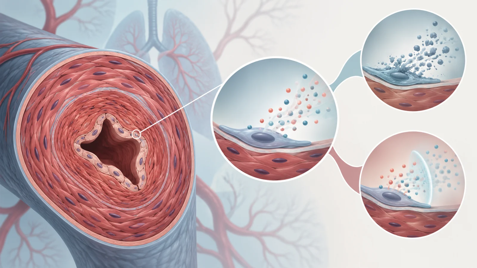
Senescent cells can release a mixture of inflammatory and growth-related substances known as the senescence-associated secretory phenotype, or SASP. Those substances can influence nearby cells, immune activity and the tissue around a blood vessel. In the pulmonary arteries, the distinction matters because a cell’s own growth arrest is different from the effects its secretions may have on neighbouring cells.
Pulmonary arterial disease begins with injury or dysfunction in the endothelial lining of the artery. Vasoactive mediators, cytokines, chemokines and growth factors can then encourage constriction, inflammation, smooth-muscle-cell proliferation and fibrosis. Excessive growth of pulmonary-artery smooth muscle cells, along with abnormal endothelial cells that resist apoptosis, thickens the arterial wall and reduces the space available for blood flow.
In experimental PH, pulmonary-artery endothelial and smooth muscle cells have been found with senescence-associated markers such as p16 and p21, alongside markers of DNA-damage responses. These observations fit a possible role for senescence in the changing vessel, but they do not by themselves show whether senescence is protecting the artery, worsening the disease or doing both at different stages.
Removing senescent cells produced opposing results
The review brings together animal experiments that point in opposite directions. In one group of studies, researchers removed senescent cells around the time pulmonary hypertension was induced. They used several approaches, including an inducible suicide gene controlled by p16, p16 gene inactivation and drugs with senolytic activity, meaning drugs intended to kill senescent cells. Removing the cells in that early setting worsened disease: more vessels became muscularised, right-ventricular systolic pressure increased and the ratio used to assess right-ventricular hypertrophy also rose.
A different rat experiment began navitoclax treatment after disease was already advanced. The rats had received monocrotaline and then an aortocaval shunt to increase pulmonary blood flow. When treatment started later, the animals showed less pulmonary vascular obstruction, lower pulmonary pressure and vascular resistance, and better measures of cardiac output and right-ventricular function. The treatment also reduced several senescence-associated markers and increased BMPR2 and Klotho in the reported analysis.
The review authors propose disease time course as one way to reconcile these findings. Senescent cells might have a protective role during an early injury response, while their persistence or secretions could become harmful once remodelling is established. That is a hypothesis about how the results may fit together, not a settled rule that early removal is harmful and late removal is beneficial.
A chronic-hypoxia mouse experiment makes the picture harder to simplify. Navitoclax was given from days 15 to 30 after hypoxia exposure, yet the animals developed worse haemodynamic and structural measures even as p16 and several inflammatory or SASP-related markers fell. A lower marker level therefore cannot be treated as proof that the disease improved. The different models, schedules and biological triggers leave timing as an important possibility, but they do not isolate it as the only cause of the opposing outcomes.
Markers show a process but not its beginning
Senescence is difficult to identify because no single universal marker defines it across tissues. A reliable assessment needs a combination of features, such as growth arrest, marker patterns, DNA-damage responses, altered cell structure and secretory changes. The review points to guidance from the NIH Cellular Senescence Network and to single-cell studies as efforts to make that identification more precise.
This matters especially when interpreting human tissue. Senescence-associated changes have been observed in pulmonary vascular tissue from people with PAH, but such samples are generally obtained at death or lung transplantation. They show what the tissue looked like at an advanced stage; they cannot, by themselves, reconstruct when senescence began, whether it initiated the vascular disease or how its role changed during progression.
The right ventricle adds another unresolved part of the story. Patients can show oxidative DNA damage, fibrosis and increased levels of several SASP-related factors in the right ventricle or blood, and the review describes evidence consistent with accelerated ageing in both lung and heart tissue. Yet defining senescence in largely non-dividing heart-muscle cells is difficult, and the molecular sequence connecting these findings to right-ventricular failure remains less clear than the vascular story.
Treatment can target the cell or its messages
Removing senescent cells is only one possible strategy. Senolytics aim to kill selected senescent cells. Senomorphics aim to reduce the SASP or alter its effects without killing the cell. A treatment that changes the signals released into the vessel may therefore have a different risk profile from one that removes the cell itself.
Preclinical studies of metformin, rapamycin and resveratrol, which have mixed antiproliferative and secretion-modulating actions, have reported beneficial effects in models of pulmonary hypertension. Those results are relevant to the senescence question, but they cannot automatically be attributed to suppression of the SASP. The same compounds affect other biological pathways, and a fall in inflammatory signals is not the same thing as proof that a particular senescent population caused the disease.
There is also a separate treatment interaction to consider. Drugs designed to stop excessive growth in pulmonary-artery cells can themselves leave surviving cells with senescence-associated features. In one experiment, barasertib, which blocks Aurora kinase B and interrupts cell division, improved pulmonary hypertension and increased the proportion of p21-positive smooth muscle cells. UC2288, a p21 attenuator, did not significantly reverse the disease on its own. Combining UC2288 with barasertib produced a greater improvement in vascular remodelling, pulmonary haemodynamics and right-ventricular function than barasertib alone.
The result does not identify a universal best combination. It shows why the biological effect of a treatment can depend on what accompanies it. For a therapy that restrains abnormal vascular growth, inducing a senescence-like state may be useful for a time, harmful if secretory activity persists, or altered again by a second drug. Selecting which cells to remove, which signals to reduce and when to intervene may matter more than treating senescence as one uniform target.
What this could change
A workable senescence-directed treatment could eventually make it possible to reduce the obstructive remodelling of pulmonary arteries more durably while easing the workload imposed on the right heart in people with PAH. Reaching that possibility would first require identifying the cell populations whose removal or signal suppression is beneficial, preserving the vascular repair that may be needed after injury, and showing in suitable studies that the intervention improves disease rather than merely changing its markers. Only then could the approach be tested and validated in patients.
The review presents that as a contemporaneous possibility, not an established clinical benefit. The central challenge is to preserve whatever protective role senescence may have during repair while preventing the persistent cellular and secretory activity that could help maintain vascular obstruction.
Key Sources
- Primary source: Cellular senescence in adult pulmonary hypertension: current state and future challenges.
DOI or Journal Page · Review or Meta-Analysis · 2025-07-01
Keep exploring