
Research Signal – Source date 2026-03-23
Type 2 diabetes is usually described through familiar mechanisms: insulin resistance, excess metabolic load and declining function of pancreatic β-cells, the cells that produce insulin. A review published on March 23, 2026, considers whether cellular senescence adds another layer to that picture.
Senescent cells are stressed cells that have entered a durable state of growth arrest. They stop dividing, but they do not necessarily become biologically silent. Instead, some release a mixture of inflammatory and tissue-altering molecules known as the senescence-associated secretory phenotype, or SASP. The review argues that this activity could connect metabolic stress with dysfunction in several organs.
The idea is biologically plausible, but it remains a research hypothesis with uneven human support. The review is a structured narrative synthesis of preclinical and human studies identified through PubMed, Scopus and Embase from January 2010 through September 2025. It synthesizes the evidence qualitatively because the models, endpoints and clinical designs were too heterogeneous for a quantitative meta-analysis. The authors also report that the review did not use a formal risk-of-bias assessment or PRISMA-based systematic methodology, so its conclusions are interpretive and hypothesis-generating.
Senescence adds a layer to familiar diabetes biology
Senescence can be triggered by several forms of cellular strain, including telomere attrition, DNA damage, oxidative stress, mitochondrial dysfunction and oncogenic signaling. Chronological aging is one influence, but the review also describes obesity, high insulin exposure, high glucose-related stress and other metabolic pressures as possible drivers of premature senescence.
That matters because type 2 diabetes does not arise from aging alone. Senescence is presented as one mechanistic contributor interacting with established forces such as obesity, insulin resistance and glucotoxicity. The proposed relationship is therefore not that diabetes has a single hidden cause. It is that persistent cellular stress may create a self-reinforcing layer of inflammation and impaired tissue maintenance on top of existing metabolic dysfunction.
The SASP is central to this explanation. Its secreted factors can change the behavior of nearby cells, recruit immune cells and influence distant tissues. In this account, senescent cells become more than passive markers of damage. Their continued signaling may help maintain the inflammatory environment associated with metabolic disease.
Why one cell state affects several organs differently
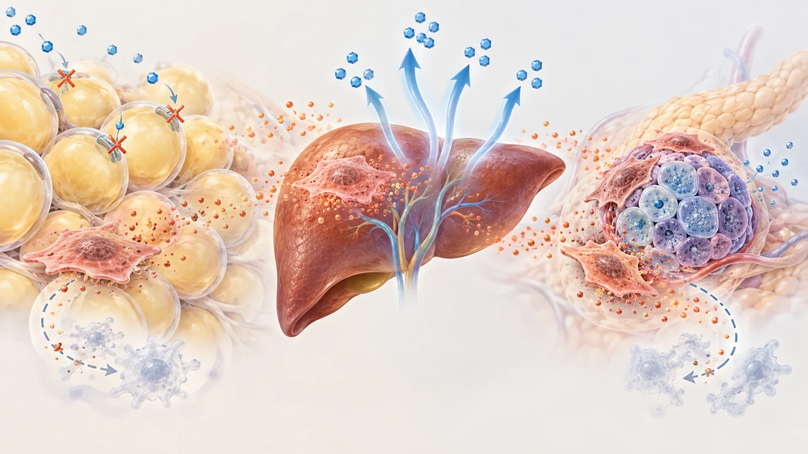
In adipose tissue, SASP-related signals can recruit and activate macrophages, alter adipocyte development and reduce GLUT4 expression. GLUT4 helps fat and muscle cells take up glucose in response to insulin. Reduced insulin-stimulated glucose uptake would make it harder for the body to clear glucose from the bloodstream.
The liver presents a different problem. Inflammation associated with senescent cells can contribute to hepatic insulin resistance and increased gluconeogenesis, the production of glucose by the liver. Excess liver glucose output is particularly relevant to fasting blood glucose, so the same broad inflammatory process can produce a different metabolic consequence depending on the tissue involved.
Pancreatic islets, which contain insulin-producing β-cells, are another setting. Chronic exposure to inflammatory signals may intensify β-cell stress, functional exhaustion and eventual cell loss. The review links these effects with disruption of insulin receptor substrate-1 signaling and with the persistent, low-grade inflammation often described as inflammaging.
This tissue-specific view is important. A senescent cell in fat, liver or an islet may share some molecular features while contributing to different failures in glucose regulation. It also explains why a treatment that changes a marker in one tissue cannot automatically be assumed to restore the whole metabolic system.
What senolytics are trying to remove
Senolytic therapies are designed to clear senescent cells by interfering with the survival systems that help those cells resist programmed cell death. These systems are called senescent cell anti-apoptotic pathways, or SCAPs. The review identifies several pathway families under investigation, including BCL-2/BCL-XL, PI3K/AKT and p53/p21/serpin signaling.
Dasatinib plus quercetin is one example of a clearance strategy discussed in the review. The aim is not simply to suppress inflammation throughout the body. It is to make selected senescent cells vulnerable to elimination while sparing healthy cells.
A related but different approach is senomorphic treatment. Rather than removing senescent cells, senomorphics seek to reduce or reshape their inflammatory secretions. The distinction matters: one strategy changes the number of senescent cells, while the other attempts to limit the signals they produce. Either approach could affect tissue behavior, but neither label by itself tells us whether glucose control or long-term disease outcomes will improve.
Mouse improvements show a mechanism, not a finished therapy
Across the animal studies summarized by the review, clearing senescent cells in adipose tissue, liver and pancreatic islets was associated with improved insulin sensitivity, reduced inflammatory burden and better preservation of β-cell function. These findings make the proposed mechanism more concrete: reducing senescent-cell activity can alter metabolic measures in living organisms.
The review includes both genetic and drug-based experiments. In an INK-ATTAC mouse model, for example, researchers used an inducible system to remove cells marked by p16Ink4a, a marker associated with senescence. Other studies used candidate senolytic drugs such as dasatinib plus quercetin, navitoclax or fisetin. Genetic removal of a marked cell population is not the same intervention as giving a drug that must reach selected tissues, interact with several pathways and avoid healthy cells.
The mouse models themselves also differ. Diet-induced obesity, leptin-deficient models and streptozotocin-induced diabetes vary in inflammatory tone, β-cell reserve and immune response. Results from one model may therefore reflect a particular form of metabolic injury rather than a universal effect across type 2 diabetes.
Exposure schedules create another translation problem. Murine studies often use intermittent, high-dose regimens, while the timing of senescent-cell clearance and possible repopulation in human metabolic tissues remains incompletely characterized. Animal results can support a mechanism worth testing, but they do not supply a clinically usable dose or prove that the same tissue changes will occur in people.
Human findings narrow the question
The human evidence summarized by the review asks more limited questions than the animal work. Small, short-duration studies involving people with type 2 diabetes and diabetic kidney disease used intermittent dasatinib plus quercetin exposure and reported reductions in senescence markers and circulating inflammatory mediators. Those findings are relevant because they suggest that the biological target can be engaged in humans.
Target engagement is still different from disease modification. A lower level of a senescence marker does not by itself demonstrate durable preservation of insulin-producing cells, restored insulin sensitivity or better long-term glucose control. In the evidence synthesized by the review, no senolytic intervention had yet demonstrated sustained improvement in HbA1c, insulin sensitivity or long-term clinical outcomes for type 2 diabetes.
The review also discusses organ-specific trials in diabetic macular edema involving UBX1325, a BCL-xL inhibitor. Those studies reported improved visual acuity with acceptable short-term safety in the summarized clinical context. Visual acuity is a meaningful patient outcome, and the eye findings prevent the human evidence from being reduced to biomarker changes alone. At the same time, an improvement in an eye-specific outcome addresses the condition of the eye, not systemic glucose regulation. It cannot establish that a senolytic treatment improves diabetes across the body.
These findings answer different questions. Animal metabolic studies ask whether clearing senescent cells can change glucose-related biology in experimental disease. Human biomarker studies ask whether treatment appears to alter the proposed cellular state or inflammatory environment. Eye studies ask whether an organ-specific senescence-related intervention can improve vision-related outcomes. Durable diabetes control would require a separate demonstration.
Selective removal is the central safety problem
Senescence is not simply a harmful identity stamped permanently onto every affected cell. It can arise as a protective response to cellular stress, limiting the division of damaged cells and contributing to tissue remodeling. A therapy that removes cells based on an imperfect marker could therefore eliminate cells whose presence still serves a useful purpose.
The challenge also includes distinguishing senescent cells from quiescent cells. Quiescent cells have stopped dividing but are not necessarily senescent. Depleting them unintentionally could damage normal tissue maintenance. The review highlights the need for more specific assays and approaches that respond to the inflammatory environments associated with harmful senescence rather than treating every nondividing cell as a target.
This makes safety a question of biological selectivity, not merely whether a drug produces side effects during a short study. Early-generation senolytics may affect survival pathways shared by healthy cells. More targeted systems are being considered, but the review does not establish a long-term dosing strategy or long-term safety profile for treating type 2 diabetes. A useful therapy would need to remove or silence the right cells, reach the relevant tissues and preserve the cells that the body still needs.
What targeting senescent cells could change for diabetes care
For people with type 2 diabetes, the potential value of this approach would be an additional treatment aimed at preserving insulin-producing cell function and supporting lasting metabolic control by reducing one contributor to tissue dysfunction. That possibility depends on more than lowering senescence markers. Human trials would need to show durable metabolic benefit while demonstrating that treatment is selective enough and safe enough for sustained use.
The review therefore places senolytics at an early translational stage. If future evidence connects selective senescent-cell targeting with preserved β-cell function, improved insulin sensitivity and clinically meaningful glucose control, the approach could add senescence-directed treatment to diabetes care. As of the review’s March 23, 2026, publication perspective, that remains a conditional possibility rather than an established treatment effect.
Key Sources
- Primary source: Cellular senescence and metabolic aging in type 2 diabetes: mechanistic insights and translational implications.
DOI or Journal Page · Review or Meta-Analysis · 2026-03-23
Keep exploring