
Research Signal – Source date 2021-05-28
A gel that heals itself sounds like a natural candidate for repairing an injury. But the two kinds of healing involve different processes. In a study published on May 28, 2021, researchers developed a water-rich gel whose temporary molecular connections could re-form after mechanical disruption. They then tested whether that material, carrying curcumin, could help create conditions for nerve repair in injured rat spinal cords.
The study connected several kinds of evidence: measurements of the gel’s strength, drug release into laboratory fluid, nerve growth in cultured tissue, and tissue and movement assessments in rats. The curcumin-loaded gel produced encouraging findings across these experiments. Understanding their significance means following the steps between a material recovering its structure and an animal recovering some function.
The biological problem is that spinal cord injury damages more than nerve fibers. Inflammation, cell death, loss of insulating myelin and cavity formation can make the injury site hostile to growth. A scaffold could give cells somewhere to attach and move, while local drug delivery could help alter that environment. Combining these jobs was the rationale for the experiment.
Filling a surgically created gap
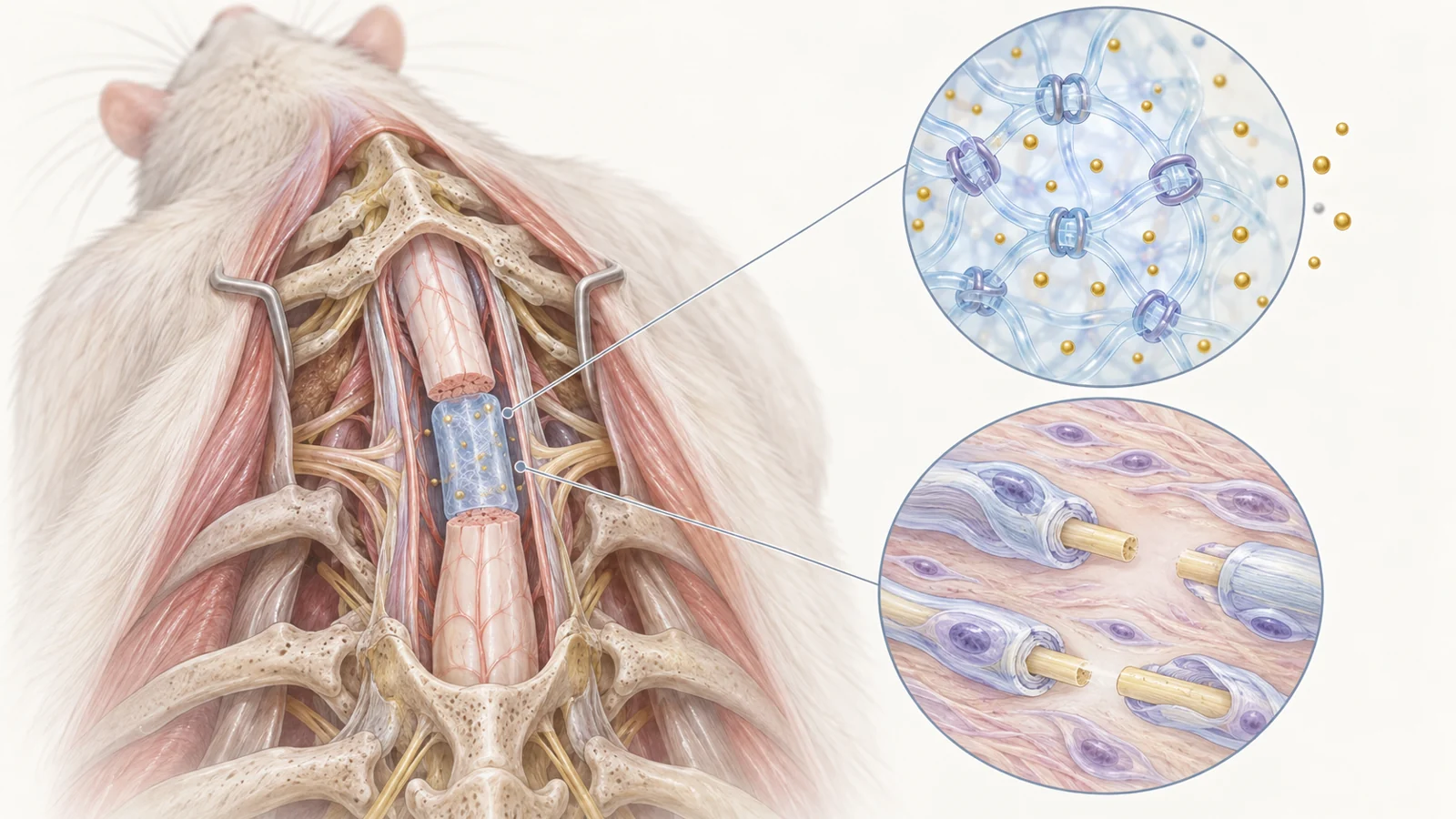
The animal study used young adult female rats. Researchers surgically exposed the spinal cord and removed a 2-millimeter segment at the ninth thoracic level, creating a complete gap. They then placed about 10 microliters of gel, or an equal volume of saline, directly into the lesion before closing the surgical opening. Treatment therefore began immediately after the injury.
Four groups allowed comparisons between saline, a modified-chitosan gel, a hybrid gel containing an added peptide, and that hybrid loaded with curcumin. The methods describe 18 rats allocated to each group, with six per group specified for the electrical and hind-limb movement assessments. Those endpoint sample sizes matter when judging how much weight to place on the functional findings.
This was a deliberately constructed lesion that could accommodate a scaffold. Although the material could pass through a needle, its use here involved an exposed spinal cord and surgery. The experiment tested local placement into a fresh injury, rather than an injection through intact skin or treatment of an established injury.
How the gel reconnects after disruption
The material’s main ingredients were chitosan modified with a chemical group called Fmoc and a peptide bearing the same group. Ring-shaped parts of these groups associate through reversible physical attractions. Together, they form numerous temporary connections that hold the water-rich network together. Adding the peptide increased the connections and strengthened the hybrid gel.
Under sufficient force, these associations can separate, allowing the material to flow. When the force subsides, they can form again. Both formulations passed through a fine, 26-gauge needle, but the hybrid emerged into laboratory buffer as an integrated strand, whereas the modified-chitosan gel formed large fragments. This gave the researchers a visible indication that the peptide changed how the material held together after injection.
Mechanical testing supplied a more quantitative measure. The researchers repeatedly alternated between strong deformation that disrupted the network and gentle deformation that allowed recovery. After five cycles, the hybrid regained approximately 88% of its storage modulus, a measure of its elastic stiffness, compared with approximately 70% for the chitosan formulation. That is the demonstrated meaning of “self-healing”: recovery of a material property after disruption. It neither measures spinal cord healing nor establishes how long the gel would retain its performance inside the body.
A week of release in laboratory fluid
Curcumin was incorporated before the gel formed. Holding a compound within a scaffold offers a way to release it gradually at a chosen location, addressing the concern that directly delivered molecules can be cleared from an injury site too quickly.
To measure release, the researchers placed gels in saline at 37 °C, gently shook them and periodically replaced the surrounding liquid. They measured curcumin in the collected fluid. Release continued over the seven-day observation period, with a faster initial phase during the first 12 hours. The authors attributed that early phase to curcumin near the gel’s surfaces, followed by slower escape from within the matrix.
The hybrid released curcumin more slowly than the modified-chitosan formulation. The authors linked this difference to the hybrid’s stronger network restricting movement of the compound. Here, “controlled release” describes how the formulation shaped release over time; it does not imply an externally adjustable dose.
These measurements establish behavior in a defined laboratory fluid. They do not determine the concentration reaching cells in an injured spinal cord, how long a useful concentration persists there, or what dose would be effective. Fluid movement and interactions with tissue make exposure inside the lesion a separate question.
Growing nerve extensions and adding insulation
The culture experiments asked whether cells could use the gel as a place to grow. Researchers placed dorsal root ganglia—clusters of sensory nerve-cell bodies—from newborn rats on the different gels. The tissue was maintained in supportive culture medium that included nerve growth factor.
Adding the peptide to the chitosan gel increased the length of neurites, the extensions growing from nerve cells. The peptide contained IKVAV, a short sequence derived from laminin, a protein associated with the matrix around cells. It was included to mimic some of laminin’s growth-supporting activity. Adding curcumin produced a further increase, with average neurite length reaching about 2 millimeters after seven days on the loaded hybrid.
The researchers also followed Schwann cells, which can wrap axons in insulating myelin. These cells migrated outward from the cultured tissue and associated with the nerve extensions, particularly on the curcumin-loaded gel. This matters because growing a long extension and equipping it to transmit signals efficiently are distinct biological tasks.
For the myelination experiments, the cultures received ascorbic acid after seven days to trigger myelin formation. After a month, staining for myelin basic protein overlapped with nerve fibers, and expression of the corresponding gene was highest on the curcumin-loaded hybrid. Together, these observations supported myelin formation under the reported culture conditions. They were not measurements of adult spinal cord circuitry, but they gave the animal experiment a specific cellular process to investigate.
What appeared inside the lesion
Two weeks after injury, the researchers examined inflammatory cells within the rat lesions. They used CD68 to identify macrophage and microglial populations and ARG1 as a marker associated with an anti-inflammatory phenotype. The curcumin-loaded hybrid had both more ARG1-positive cells and a higher proportion of ARG1-positive cells among the CD68-positive population than the other groups.
That pattern is consistent with a change in the local inflammatory response. It does not mean inflammation had simply disappeared. The measurements described which cells and markers were present, supporting the proposed change in the repair environment without proving that this change caused every later benefit.
Schwann cells also entered the graft. They were more numerous in the curcumin-loaded hybrid than in either gel without curcumin and were seen extending into it from the region of nearby nerve roots. This moved the inquiry beyond whether cells could migrate on a gel in culture to whether cells already present in the animal could populate the implanted material.
The evidence for insulation included more than the presence of a myelin-associated marker. Cross-sectional images showed myelin staining forming rings around axons. Electron microscopy revealed layered myelin surrounding axonal structures, with features consistent with Schwann cells participating in that wrapping. The loaded hybrid showed stronger myelin-related findings than the comparison groups.
These images support local remyelination within the graft. Establishing that axons have acquired insulation is still different from establishing where those axons connect, whether their connections are appropriate, and how much useful information travels through them. Those questions make functional testing essential.
Did the tissue changes translate into movement?
The study went beyond microscopy. Researchers assessed hind-limb movement using the Basso–Beattie–Bresnahan locomotor scale, which scores observed movement in an open field. They also measured motor evoked potentials at eight weeks after injury, recording electrical responses to assess motor-system conduction.
The two approaches address different questions. Movement scoring describes what the animal does, while an evoked electrical response probes how the motor system responds to stimulation. The authors reported improved hind-limb movement and interpreted the electrical findings as improved conduction, particularly with the curcumin-loaded hybrid.
There is a reason to be restrained about the electrical result. The results prose contains an inconsistency between the stated directions of changes in response amplitude and delay and the interpretation that follows. It supports reporting the authors’ overall interpretation with caution, rather than assigning a precise improvement in signal size or speed. Nor do the reported functional findings establish restoration of normal walking or complete neural circuitry.
Taken together, the material, cell, tissue and functional experiments form a plausible repair account. Their agreement strengthens the case for further investigation, but it does not independently establish each causal step from curcumin release to inflammation, myelination and movement.
What this experiment leaves unresolved
The group comparisons help show what changed when the peptide and then curcumin were added. They cannot isolate an advantage over curcumin delivered without a gel, because free curcumin was not one of the four listed animal treatments. They also do not establish synergy—the stronger claim that combining components produces more benefit than their separate effects would predict. Changes in structure, cell interactions and drug delivery all contribute possible explanations.
Translation also depends on the injury being treated. An immediately filled surgical gap in young adult female rats is a specific setting. Effectiveness in chronic injuries, in tissue with different patterns of damage, or across a wider range of animals remains a separate question. The culture work adds another distinction, because newborn sensory tissue in growth-supporting medium differs from an adult injured spinal cord.
For use in people, the material would need evidence of safe behavior over time, reliable delivery at a suitable dose, and meaningful functional benefit in relevant injury settings. The experiments here do not establish human therapeutic benefit or safety. Those are substantial requirements beyond showing favorable cells and tissue inside a graft.
What this could change
If the approach produces durable, useful nerve connections and its delivery and safety problems can be solved, a lesion-filling gel could make local treatment do two jobs at once. It could provide a temporary place for repair-associated cells and axons to organize while keeping a drug near the damaged tissue during a period when the local environment matters.
The potential clinical payoff would be a way to support recovery at the injury site with a material that conforms to the available space and releases its cargo gradually. If the resulting axons form appropriate connections and transmit useful signals, that could contribute to recovery of movement after spinal cord injury. The study offers an experimental basis for pursuing that possibility, with the eventual value depending on functional repair that lasts beyond the early response to implantation.
Key Sources
- Primary source: An injectable and self-healing hydrogel with controlled release of curcumin to repair spinal cord injury.
DOI or Journal Page · Peer-Reviewed Paper · 2021-05-28
Keep exploring