
Research Signal – Source date 2024-07-31
A protective coating on a device that contacts blood has more than one job. Resisting clot formation initially is valuable, but maintaining that protection as the surface becomes damaged is a separate challenge. Inflammation adds another demand. A research article published on July 31, 2024, describes an experimental polymer coating designed to address all three through a combination of an anti-clotting drug, repair of coating damage and two components intended to control inflammation.
The authors report clot prevention and inflammation modulation in laboratory and in vivo experiments. Their proposal brings several functions into one material, with different ingredients incorporated in different ways. Understanding that construction helps explain both the attraction of the approach and why its promise of lasting protection needs careful interpretation.
Building the anti-clotting drug into the material
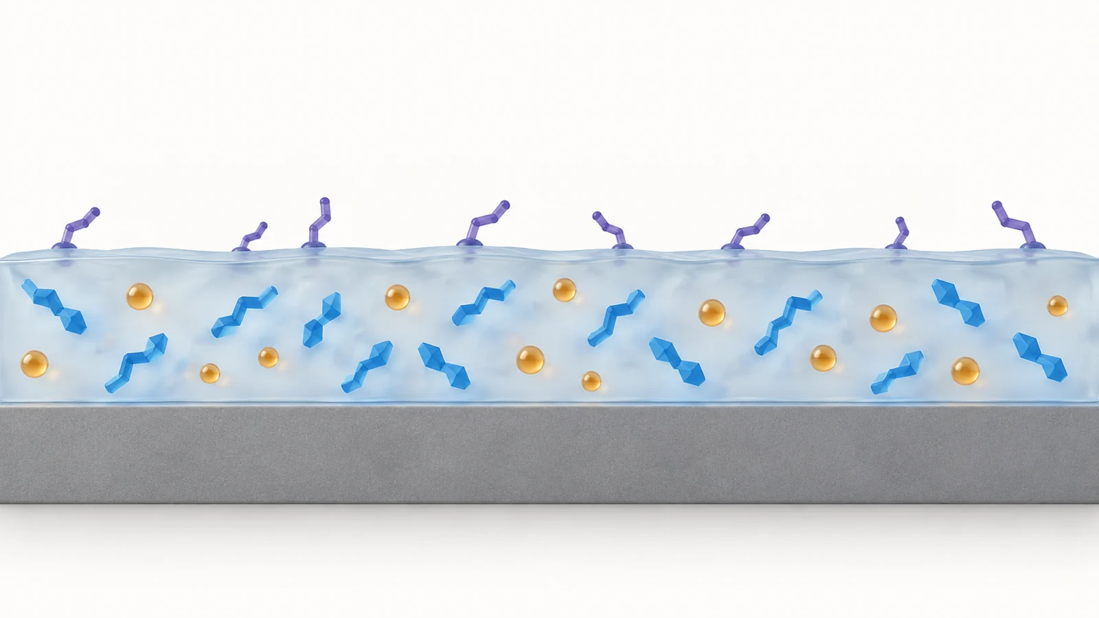
Heparin is an anti-clotting drug. In this coating, the researchers chemically modify it with methacrylate groups, producing a form they call MA-heparin. They then combine it with methyl methacrylate and n-butyl acrylate, two small molecules used to make the polymer. Heat initiates the chemical reaction that joins these ingredients during polymer formation.
That detail matters because heparin participates in making the material. Describing a surface simply as “heparin-coated” would leave out how the drug becomes incorporated. The authors report that this approach increases the coating’s heparin content and inhibits thrombosis, the formation of blood clots.
They also describe the resulting coating as self-healing, meaning that it can repair damage to the coating itself. The term refers to the material covering the device; it does not mean that the device’s underlying structure or a patient’s tissue repairs itself. Incorporating heparin and achieving self-healing are reported features of the formulation, but the abstract alone does not explain the physical process responsible for repair.
Two different ways to address inflammation
The inflammation-related ingredients enter the coating through different routes. One is a polymer called PVAX, a polyoxalate containing vanillyl alcohol. After the initial coating is formed, the researchers chemically attach PVAX through a reaction called esterification. They describe it as responsive to reactive oxygen species, or ROS—chemically reactive oxygen-containing substances—and assign it a role in countering acute inflammation.
Aspirin is introduced earlier. It dissolves in the mixture of methyl methacrylate and n-butyl acrylate and becomes encapsulated as the coating forms. The authors describe its subsequent release as temperature-dependent and associate it with control of chronic inflammation.
These are distinct response dependencies. PVAX responds to a chemical condition involving reactive oxygen species; aspirin release depends on temperature. Neither description, by itself, establishes an automatic treatment schedule in which one ingredient acts first and the other takes over later. The acute and chronic roles express the authors’ account of the components’ functions, while the timing and coordination of those functions require experimental detail.
How much protection do the reported results establish?
The article’s statement of significance reports application to varied substrates, along with demonstrations of inflammation modulation and thrombosis prevention in vitro and in vivo. Those findings support interest in the coating as an experimental material. Applying it to different surfaces also matters for a strategy intended for devices made from different materials.
This account is based on the PubMed abstract and statement of significance. They do not provide enough detail to assess the experimental models, sample sizes, comparison groups, effect sizes or follow-up periods. Consequently, the reported results cannot be translated here into a percentage reduction in clotting or a duration of protection. The authors’ language about long-term stability does not, on this basis, establish how long a coated device could function in use.
Repair also has several dimensions. The conditions needed for healing, its speed and its performance after repeated damage cannot be established from this summary. Closing a damaged region and restoring its anti-clotting or inflammation-related functions are different questions. Assessing the latter would require evidence that those particular functions recover after damage.
Nor does the available summary establish a specific in vivo model or outcomes in patients. It cannot support conclusions about bleeding risk, aspirin exposure in clinical use or a reduced need for systemic anticoagulation. Manufacturing at scale and tolerating sterilization are further requirements for practical deployment that cannot be assessed here. These are limits of what can be verified from the abstract, rather than assertions that the full paper omits those investigations.
What this could change
If the coating can retain its protective functions after damage and its inflammation-related components work under the conditions of device use, it could provide a way to maintain a more blood-compatible surface throughout use. Incorporating clot inhibition, material repair and inflammation control into the same coating could then help reduce complications arising where blood meets a device. Reaching that benefit depends on demonstrating durable performance and acceptable safety in the relevant setting, but the research offers a concrete material design through which those capabilities might be combined.
Key Sources
- Primary source: Heparinized self-healing polymer coating with inflammation modulation for blood-contacting biomedical devices.
DOI or Journal Page · Peer-Reviewed Paper · 2024-07-31
Keep exploring