
Research Signal – Source date 2021-11-15
A cancer-fighting immune response can create an unexpected problem. Signals released by immune cells may help them attack a tumor, yet the same signals can prompt prostate cancer cells or nearby immune-regulating cells to increase PD-L1, a protein that can restrain immune activity through contact with PD-1. The tumor is not simply hiding from an immune response that never arrived. In some circumstances, it may be adapting to one that is already underway.
That tension is the subject of a systematic literature review published on November 15, 2021. The review followed PRISMA guidelines and examined how the prostate cancer tumor microenvironment and intracellular signaling pathways influence the PD-1/PD-L1 axis. Its evidence included human and mouse cell-line experiments, animal models and observations involving human tumors or patient-associated material. These forms of evidence answer different questions, so the mechanisms are more informative than a simple promise that blocking PD-L1 will work for every patient.
When an immune alarm raises a brake
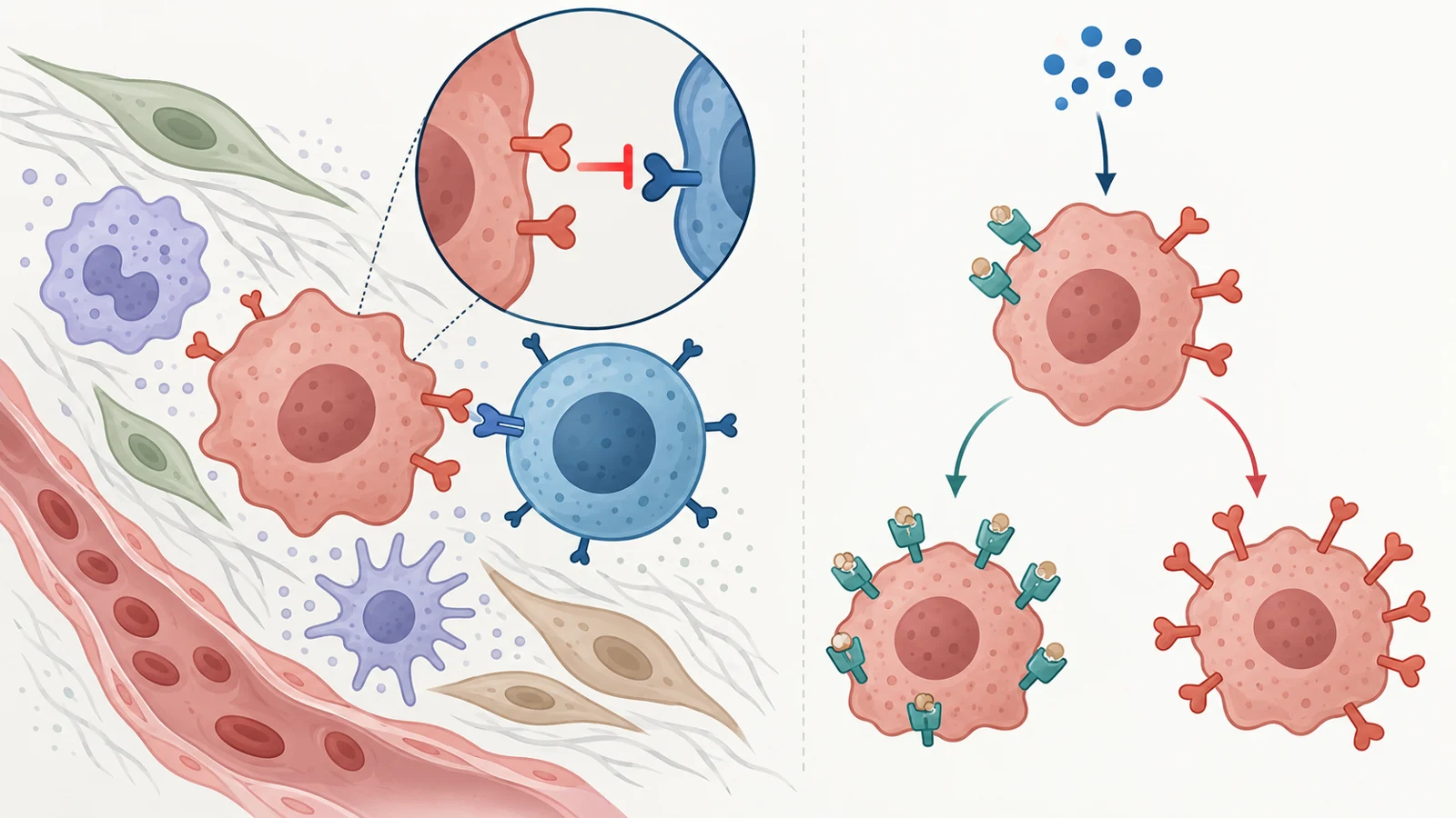
Interferon-gamma is a useful place to see the problem. CD8-positive T cells, natural killer cells and macrophages release it to support immune-cell activity, antigen processing and the development of tumor-infiltrating lymphocytes. In responsive prostate cancer cell lines, however, interferon-gamma also increased PD-L1 through the JAK/STAT pathway. Once increased, PD-L1 could suppress the activity and proliferation of cytotoxic T cells and natural killer cells, and in the reviewed experiments it was associated with T-cell apoptosis.
The same pathway can influence both sides of immune recognition. Interferon-gamma increased PD-L1 and HLA-ABC, part of the major-histocompatibility-complex class I machinery that helps display non-self targets to CD8-positive T cells. A prostate cancer cell can therefore show more of the molecular material that helps immune cells recognize it while also increasing a signal that limits the attack. The result is not a tidy switch from immune visible to immune invisible. It is a biological contest between recognition and restraint.
The response was not uniform across the models. Much of the interferon-gamma work involved metastatic, androgen-insensitive cell lines, and the review also describes lines that were insensitive to the cytokine. Alterations in JAK/STAT signaling, including loss of JAK1 expression in LNCaP cells, were reported in some insensitive lines. A signaling pathway that raises PD-L1 in one laboratory model therefore cannot automatically be treated as a universal treatment target. Blocking it might change several immune-related processes at once, including processes involved in presenting tumor targets.
The neighborhood can keep immune cells at the edge
The tumor microenvironment changes more than PD-L1 levels. Prostate cancer was described in the review as frequently having sparse lymphocytic infiltration or T-cell exclusion, with T cells remaining in adjacent stromal or benign tissue rather than making direct contact with cancer cells. Impaired antigen presentation can further limit T-cell activation. Even when tumor-specific CD8-positive T cells are present, they may show signs of exhaustion, including PD-1 and Tim-3 expression.
This makes access and coordination as important as the presence of an immune cell. Tumor and surrounding suppressive cells can express PD-L1, and an increase in PD-L1 may accompany a productive antitumor response rather than appear only in a completely inactive tumor environment. The surrounding tissue can also shape which immune cells arrive, how they differentiate and whether they can present tumor antigens effectively.
The review gives a concrete example involving stromal cells, CCL2 and IL-6. In the cited experimental work, prostate cancer stromal cells released CCL2, which attracted monocytes. CCL2 also activated CD11b-positive monocytes and increased their IL-6 expression. IL-6 favored PD-L1 upregulation, while also increasing local CCL2 levels, creating a feedback loop linked to monocyte infiltration, malignant-cell proliferation and tumor survival.
Stromal signals affected dendritic cells as well. Stromal factors and IL-6 influenced their differentiation through STAT3-related activity. Dendritic cells generated under these conditions showed higher PD-L1, produced more IL-10 and less IL-12 after stimulation, and were less able to cross-present tumor antigens to CD8-positive T cells. They also suppressed T-cell proliferation. The important change here is not only a stronger brake on an individual T cell. It is a reshaped neighborhood in which immune cells may be recruited, instructed and positioned in ways that make effective recognition harder.
Treatment can change the immune neighborhood
The environment was not fixed in the reviewed studies. In prostate cancer mouse models, amphiregulin released by treatment-damaged stroma increased PD-L1 on cancer cells and contributed to resistance to immune surveillance. The finding links damage in surrounding tissue with a change in tumor-cell immune regulation within those models. It does not show that chemotherapy or radiotherapy generally harms patients through this mechanism, but it does show why the timing and context of treatment can matter biologically.
Another mouse experiment tested a different kind of intervention. In TRAMP-C2 prostate cancer models, IL-15 was combined with antibodies blocking CTLA-4 and PD-L1. The combination increased the cytotoxic activity of CD8-positive T cells and antigen-specific interferon-gamma release, reduced suppressive regulatory-T-cell activity and decreased tumor growth while prolonging survival in the tumor-bearing animals.
The cell-depletion results helped identify what was necessary for that experimental response. Removing CD8-positive cells eliminated the antitumor effects of IL-15 and the combination treatment. Although IL-15 expanded natural-killer-cell numbers, depleting NK1.1-positive cells did not change tumor growth in that experiment. This does not establish that natural killer cells are unimportant in prostate cancer generally. It shows that, in that model and treatment setting, the CD8-positive response was essential to the observed effect.
The review also notes a safety concern with IL-15. IL-15 responses could produce high levels of inflammatory cytokines, including IL-6, IL-10, tumor necrosis factor alpha and interferon-gamma, which may lead to serious side effects in patients. A combination that improves immune-cell activity in an animal model therefore still has to be translated into a tolerable human treatment, with its benefits and risks tested rather than assumed.
What the experiments can and cannot tell us
The intracellular pathway evidence was unevenly distributed. ERK/MEK and Akt-mTOR were studied in human metastatic androgen-independent cell lines such as PC3 and DU145. NF-kB and WNT were also investigated in LNCaP cells, while JAK/STAT was examined across a broader set of human lines and in a mouse line. Across the reviewed experiments, activation of ERK, NF-kB and JAK/STAT was associated with increased PD-L1, and inhibitors of MEK, JAK or STAT reduced PD-L1 expression in the tested prostate cancer cells. PTEN-related inhibition of mTOR and PD-L1 also reduced tumor growth in a preclinical mouse setting involving chemerin.
These findings contain several different kinds of result. A change in PD-L1 is a molecular observation. A reduction in T-cell or natural-killer-cell function is a functional immune observation. Smaller tumors or longer survival in mice are animal outcomes. None of those findings, by itself, establishes that PD-L1 expression alone can select an effective treatment for an individual patient, or that the highlighted mouse combination improves survival in people with prostate cancer.
The variation between cell lines matters as well. Some were sensitive to interferon-gamma and some were not. Certain pathways and extracellular factors were examined repeatedly, while others received less attention. The review therefore offers a set of connected mechanisms rather than a single universal prostate cancer pathway. It also does not establish a preferred clinical combination, a patient benefit-to-risk balance or a magnitude of benefit.
From the November 2021 perspective, the practical question was how to use this complexity. Blocking PD-1/PD-L1 signaling could restore some antitumor immune activity by improving T-cell activation and cytotoxic killing, but the surrounding microenvironment, the cell’s internal signaling state and treatment-related changes may all influence the result. Understanding those relationships could help explain why some tumors resist immunotherapy, without turning one laboratory measurement into a guaranteed clinical answer.
How this could broaden the benefit of prostate cancer immunotherapy
Patients whose tumors resist immune treatment could eventually benefit from combinations that restore cancer-killing immune activity while addressing a demonstrated source of suppression, such as a pathway or surrounding-cell interaction that raises PD-L1. The conditional opportunity is to make immunotherapy useful for more people by matching checkpoint blockade with the biological obstacle that is limiting immune access or function. That possibility depends on translating these mechanisms into patient benefit without unacceptable toxicity; the reviewed experiments had not yet established the necessary clinical combination or outcome.
Key Sources
- Primary source: What Do We Have to Know about PD-L1 Expression in Prostate Cancer? A Systematic Literature Review. Part 3: PD-L1, Intracellular Signaling Pathways and Tumor Microenvironment.
DOI or Journal Page · Review or Meta-Analysis · 2021-11-15
Keep exploring