
Research Signal – Source date 2021-02-19
A study published on February 19, 2021, found that people with Parkinson’s disease who performed worse on a cognitive screening test also tended to show weaker slow-wave activity during sleep. The relationship was not spread evenly across the head or across all wave frequencies. It was strongest in recordings from the front of the head and most pronounced for waves cycling one to two times per second.
The finding offers a more specific clue than simply asking how long someone slept or how much time they spent in different sleep stages. It also remains an association observed at one point in time. The study cannot tell whether weaker slow waves contribute to cognitive problems, whether cognitive problems affect sleep, or whether both reflect other aspects of Parkinson’s disease.
The study looked beyond how long people slept
The researchers analyzed 140 people with Parkinson’s disease who did not have a clinical diagnosis of dementia. They came from a standardized work-up used to assess eligibility for deep brain stimulation between January 2012 and July 2018. Cognitive testing and sleep recording were part of that work-up regardless of whether a participant had reported sleep or thinking difficulties.
Cognitive performance was assessed with the Montréal Cognitive Assessment, commonly called the MoCA. For descriptive purposes, the researchers classified scores below 26 as a positive screen for cognitive impairment and scores of 26 or higher as a negative screen. Thirty-five participants screened positive and 105 screened negative. Those numbers describe this selected clinical sample; they are not an estimate of how common cognitive impairment is among everyone living with Parkinson’s disease.
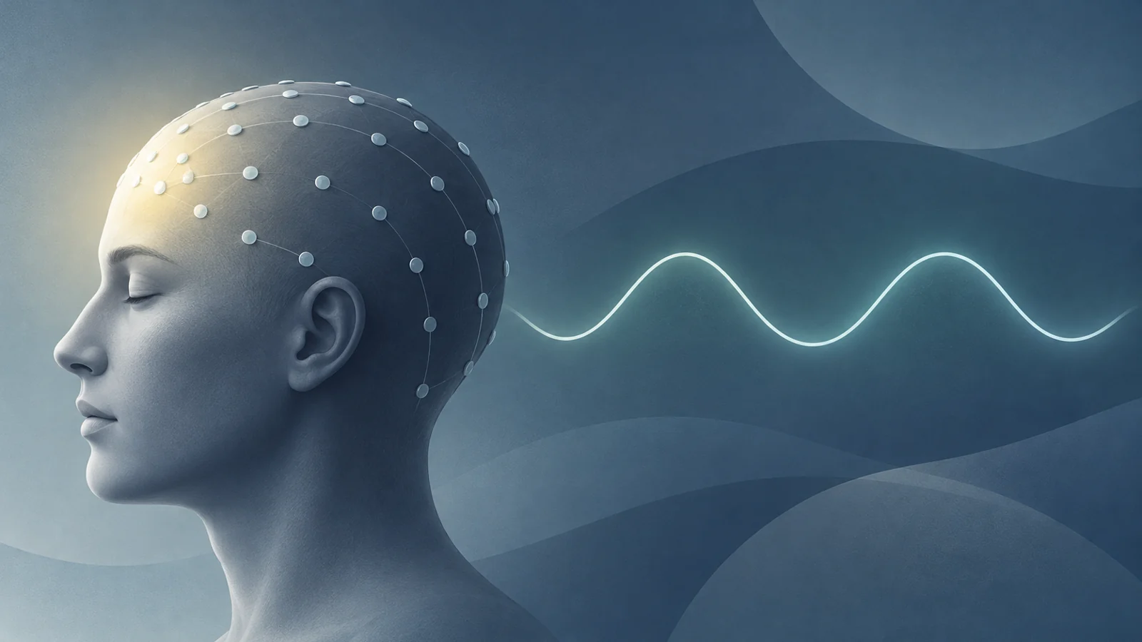
Each participant also underwent one night of video-polysomnography in a sleep laboratory. Sleep experts scored the night’s sleep stages, including non-rapid eye movement sleep, or NREM sleep, and the researchers analyzed electrical activity recorded from frontal, central and occipital locations. The analysis focused on N2 and N3 sleep, stages in which slow activity could be measured across the night.
What the sleep recording measured
Slow-wave activity describes the strength of slow electrical rhythms in the sleeping brain. It is different from the number of minutes a person spends asleep, the percentage of the night spent in a particular stage, or the presence of a sleep disorder. A person could therefore have broadly similar conventional sleep measures to another participant while showing a different distribution of electrical activity within those stages.
The researchers divided the recorded activity into frequency bands. Their main slow-wave range was 1–4 Hz, meaning electrical cycles occurring roughly one to four times per second. They also separated that range into narrower bands, including 1–2 Hz, 2–3 Hz and 3–4 Hz. The 1–2 Hz band was important because it allowed the analysis to ask whether the slowest part of the broader range carried the clearest relationship with cognitive performance.
Rather than relying only on the overall size of a person’s recording, the primary analysis measured each band’s relative share of total recorded power during N2 and N3 sleep. The models also considered absolute power as an additional analysis. Age, sex, disease duration, education, motor phenotype, levodopa-equivalent dose and the total number of sleep medications were included as statistical controls.
Conventional polysomnography characteristics did not differ between the participants who screened positive and those who screened negative. Looking at the finer-grained electrical pattern therefore added information that was not apparent from the broader sleep-stage measures alone.
The strongest pattern was near the front
Poorer MoCA performance was associated with reduced regional slow-wave activity in the 1–4 Hz range. The result was driven mainly by the 1–2 Hz component rather than by the higher frequencies within the broader delta range. In practical terms, the relevant difference was not simply less sleep. It was a lower relative contribution of 1–2 Hz activity during NREM sleep.
Location mattered. The association followed a front-to-back gradient: it was strongest over frontal recordings, weaker over central recordings and not detected over occipital recordings in this analysis. The reported correlation was rho = 0.33 for the frontal recordings and rho = 0.28 for the central recordings, both with p < 0.001. No corresponding association was detected at the occipital locations.
A correlation describes how two measurements varied together across a group. It does not provide a threshold that can classify one person. A participant with a particular sleep recording cannot be assigned a cognitive diagnosis or a future outcome from these correlation values alone.
The regional result also helps explain why studies of slow-wave activity and cognition can produce different findings. A measurement averaged across the whole scalp could blur a relationship that is concentrated in one area. The researchers discussed earlier Parkinson’s studies with inconsistent results, including one smaller study that did not find an association with NREM delta power and another that linked frontal slow-wave activity with global cognition. In this study, the larger sample and the separation of recordings by location and one-hertz frequency bands made the pattern more specific, while still leaving its meaning unresolved.
A screening score is not a diagnosis
The study’s cognitive result concerns performance on a screening measure, not a dementia diagnosis. Participants with a clinical diagnosis of dementia were excluded, and the researchers used the MoCA cutoff to describe who screened positive or negative within the sample. A positive screen can indicate that more detailed assessment may be relevant, but it does not establish the cause, severity or future course of an individual’s cognitive difficulty.
The study was retrospective and cross-sectional. The researchers examined information already collected during a clinical work-up and compared sleep recordings with cognitive performance at that time. They did not follow participants to determine whether weaker frontal slow-wave activity predicted later decline. The paper’s result therefore supports a concurrent relationship, not a forecast.
The sample also narrows how far the finding can be generalized. These participants were evaluated in a specialized pathway connected with deep brain stimulation eligibility, so they may not represent the full range of people with Parkinson’s disease. The recording came from a single laboratory night, which allows a possible first-night effect. Sleep and cognition can also be influenced by factors that were not systematically measured in the analysis, including depression, nocturia and nighttime changes in Parkinson’s symptoms.
Medication differences complicate the picture further. Participants with cognitive impairment more often took several medications that could affect sleep or cognition, and those medications were also associated with less slow-wave activity. Adjusting for medication effects did not remove the reported association between MoCA performance and slow-wave activity, but the authors still called for more detailed study of how medication, sleep and cognition interact.
Most importantly, the design cannot determine direction. Reduced slow-wave activity might accompany cognitive impairment, cognitive impairment might accompany changes in sleep, or both might be shaped by shared disease processes. The study also does not show that increasing slow-wave activity would improve cognition or prevent dementia.
How sleep recordings could help assess cognitive vulnerability
The finding points toward a possible future use of sleep recordings as a physiological companion to cognitive testing. A measure that captures regional and frequency-specific sleep activity could eventually help clinicians characterize cognitive vulnerability in people with Parkinson’s, especially if it provides information that ordinary sleep-stage measures and existing assessments do not capture.
That possibility depends on evidence the study did not provide. Larger samples would need to test whether the pattern holds across different clinical populations, and longitudinal research would need to establish whether it relates to later cognitive trajectories. Researchers would also need to show that the sleep measure adds useful information beyond established cognitive assessment and can be interpreted reliably for individual patients. Until then, the result is a promising observational association, not a ready diagnostic test or a reason to pursue a sleep treatment.
Key Sources
- Primary source: Reduced Regional NREM Sleep Slow-Wave Activity Is Associated With Cognitive Impairment in Parkinson Disease.
DOI or Journal Page · Peer-Reviewed Paper · 2021-02-19
Keep exploring